You have no items in your shopping cart.
Cart summary
Item 1 of 5
Item 1 of 5
LATS1 Antibody
Catalog Number: orb443195
| Catalog Number | orb443195 |
|---|---|
| Category | Antibodies |
| Description | LATS1 Antibody |
| Species/Host | Rabbit |
| Clonality | Polyclonal |
| Tested applications | ELISA, FC, ICC, IHC, IHC-Fr, WB |
| Reactivity | Human, Mouse, Rat |
| Isotype | Rabbit IgG |
| Immunogen | E. coli-derived human LATS1 recombinant protein (Position: Q637-A698). |
| Concentration | Adding 0.2 ml of distilled water will yield a concentration of 500 μg/ml. |
| Dilution range | Western blot, 0.1-0.5μg/ml Immunohistochemistry (Paraffin-embedded Section), 0.5-1μg/ml Immunohistochemistry (Frozen Section), 0.5-1μg/ml Immunocytochemistry, 0.5-1μg/ml Flow Cytometry, 1-3μg/1x106 cells Direct ELISA, 0.1-0.5μg/ml |
| Form/Appearance | Lyophilized |
| Conjugation | Unconjugated |
| MW | 150 kDa |
| UniProt ID | O95835 |
| Storage | Store at -20˚C for one year from date of receipt. After reconstitution, at 4˚C for one month. It can also be aliquotted and stored frozen at -20˚C for six months. Avoid repeated freeze-thaw cycles. |
| Alternative names | Serine/threonine-protein kinase LATS1; Large tumor Read more... |
| Note | For research use only |
| Application notes | Add 0.2ml of distilled water will yield a concentration of 500ug/ml. |
| Expiration Date | 12 months from date of receipt. |
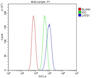
Flow Cytometry analysis of SiHa cells using anti-LATS1 antibody (Blue line).Isotype control antibody (Green line) was rabbit IgG .Unlabelled sample (Red line) was also used as a control.
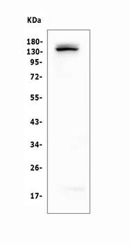
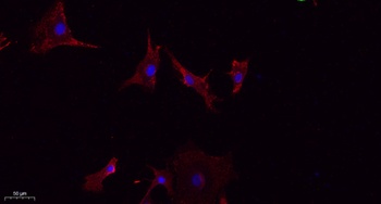
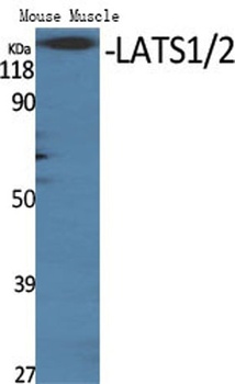
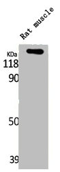
WB analysis of LATS1 using anti-LATS1 antibody.Lane 1:human K562 Cell.
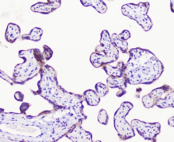
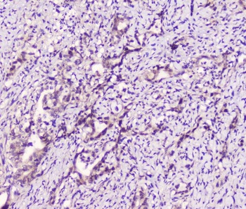
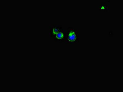
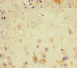
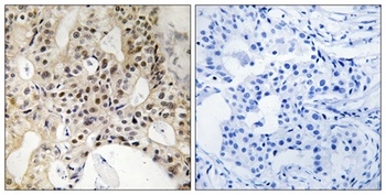
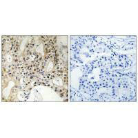
IHC analysis of LATS1 1using anti-LATS1 antibody.LATS1 was detected in paraffin-embedded section of human placenta tissue.
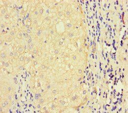
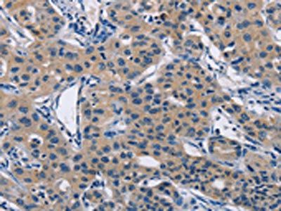
IHC analysis of LATS1 using anti-LATS1 antibody.LATS1 was detected in paraffin-embedded section of human cholangiocarcinoma tissue.
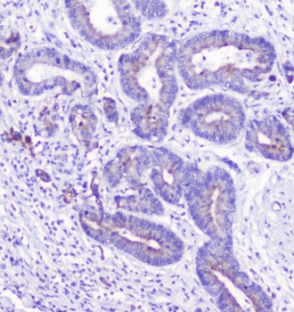
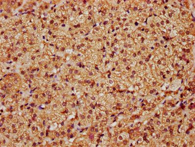
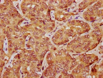
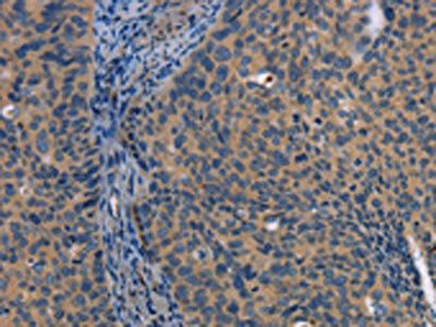
IHC analysis of LATS1 using anti-LATS1 antibody.LATS1 was detected in paraffin-embedded section of human rectal cancer tissue.
- Item 1 of 5
- Item 1 of 4
- Item 1 of 2
- Item 1 of 1
- Item 1 of 1

Submit a review
Filter by Rating

- 5 stars

- 4 stars
- 3 stars
- 2 stars
- 1 stars