You have no items in your shopping cart.
Cart summary
Item 1 of 3
Item 1 of 3
KRT10 Antibody / Cytokeratin 10
Catalog Number: orb2637817

| Catalog Number | orb2637817 |
|---|---|
| Category | Antibodies |
| Description | This antibody recognizes a protein of 56.5kDa, identified as cytokeratin 10 (CK10). cytokeratin 10 is expressed in all suprabasal layers of the epidermis. In the epidermis, expression of cytokeratin 10 strictly parallels the extent of differentiation; it is absent in the basal layer, appears in the first suprabasal layers and increases in concentration towards the granular layer. However, cytokeratin 10 is rarely detected in early stages of vulvar squamous carcinomas (tumors less than 2 cm, clinical stage I) regardless of the tumor grade. In larger and more advanced tumors (greater than 2 cm, clinical stages II and III), cytokeratin 10 is detected very frequently. Expression of cytokeratin 10 is related to maturation of malignant keratinocytes, being preferentially detected in more-differentiated parts. |
| Species/Host | Mouse |
| Clonality | Monoclonal |
| Clone Number | LH2 |
| Tested applications | IHC-P, WB |
| Reactivity | Human, Mouse |
| Isotype | Mouse IgG1, kappa |
| Immunogen | Skin extract from a human psoriasis patient was used as the immunogen for this Cytokeratin 10 antibody. |
| Antibody Type | Primary Antibody |
| Dilution range | Western blot: 1-2ug/ml,Immunohistochemistry (FFPE): 0.5-1ug/ml for 30 min at RT |
| Purity | Protein G affinity chromatography |
| Conjugation | Unconjugated |
| Formula | 0.2 mg/ml in 1X PBS with 0.1 mg/ml BSA (US sourced) and 0.05% sodium azide |
| Hazard Information | This Cytokeratin 10 antibody is available for research use only. |
| Entrez | 3858 |
| Storage | Maintain refrigerated at 2-8°C for up to 2 weeks. For long term storage store at -20°C in small aliquots to prevent freeze-thaw cycles. |
| Buffer/Preservatives | 0.2 mg/ml in 1X PBS with 0.1 mg/ml rAlbumin (US sourced) and 0.05% sodium azide |
| Note | For research use only |
| Application notes | The concentration stated for each application is a general starting point. Variations in protocols, secondaries and substrates may require the antibody to be titered up or down for optimal performance.1. Staining of formalin-fixed tissues requires boiling tissue sections in 1mM EDTA, pH 8-9, for 10-20 min followed by cooling at RT for 20 minutes.2. The prediluted format is supplied in a dropper bottle and is optimized for use in IHC. After epitope retrieval step (if required), drip mAb solution onto the tissue section and incubate at RT for 30 min. |
| Expiration Date | 12 months from date of receipt. |

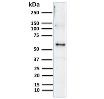
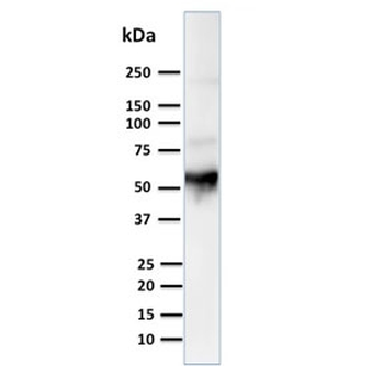
Western blot testing of human thymus lysate with Cytokeratin 10 antibody. Predicted molecular weight ~59 kDa.

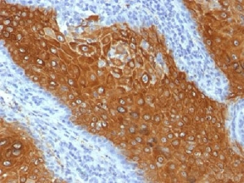
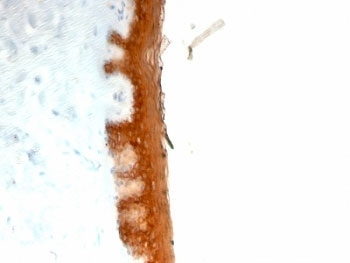
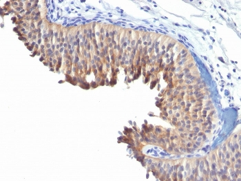
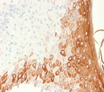
IHC testing of human bladder carcinoma with Keratin 10 antibody (clone LH2).

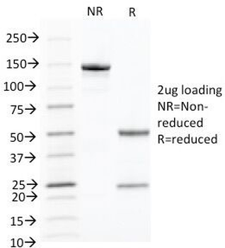
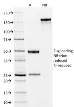
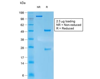
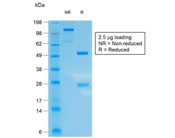
SDS-PAGE analysis of purified, BSA-free Cytokeratin 10 antibody (clone LH2) as confirmation of integrity and purity.
- Item 1 of 4
- Item 1 of 4
KRT10 Antibody / Cytokeratin 10 [orb2638754]
IHC-P, WB
Human, Mouse
Mouse
Monoclonal
Unconjugated
100 μg - Item 1 of 3
- Item 1 of 2
- Item 1 of 2