You have no items in your shopping cart.
Cart summary
Item 1 of 9
Item 1 of 9
JDP2 Antibody
Catalog Number: orb1733245
| Catalog Number | orb1733245 |
|---|---|
| Category | Antibodies |
| Description | JDP2 Antibody |
| Species/Host | Rabbit |
| Clonality | Recombinant |
| Tested applications | FC, ICC, IHC, WB |
| Reactivity | Human, Mouse |
| Immunogen | between 50 and 100 |
| Concentration | 400 µg/ml |
| Dilution range | WB: 1:1000, IHC: 1:40 to 1:100. Epitope retrieval with Tris-EDTA pH 9.0 is recommended for FFPE tissue sections, ICC: 1:40 to 1:100. Epitope retrieval with Tris-EDTA pH 9.0 is recommended for FFPE cell sections |
| Form/Appearance | Whole IgG |
| Conjugation | Unconjugated |
| Target | JDP2 |
| UniProt ID | Q8WYK2 |
| Storage | Shelf life: 1 year from date of receipt. Storage: 2 - 8°C |
| Buffer/Preservatives | Borate Buffered Saline (BBS) pH 8.2 with 0.1% rAlbumin and 0.09% Sodium Azide |
| Alternative names | JUNDM2; progesterone receptor co-activator; jun di Read more... |
| Note | For research use only |
| Application notes | Format: Whole IgG |
| Expiration Date | 12 months from date of receipt. |

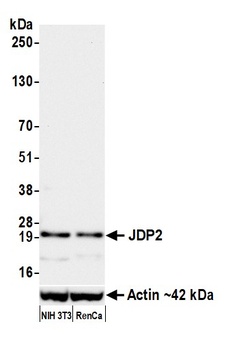
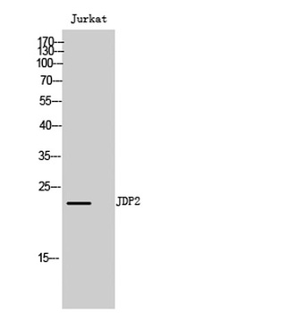
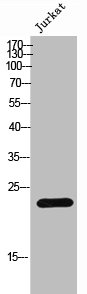
Detection of mouse JDP2 by western blot. Samples: Whole cell lysate (50 µg) from NIH 3T3 and RenCa cells prepared using NETN lysis buffer. Antibody: Rabbit anti-JDP2 recombinant monoclonal antibody (orb1733245) used at 1:1000. Secondary: HRP-conjugated goat anti-rabbit IgG. Detection: Chemiluminescence with an exposure time of 30 seconds. Lower Panel: Rabbit anti-Actin recombinant monoclonal antibody.

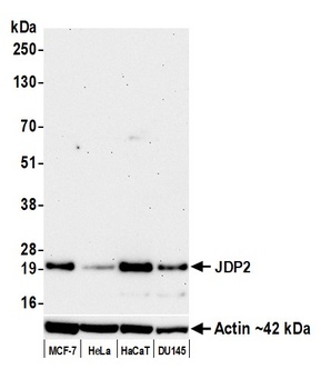
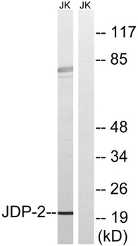
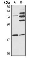
Detection of human JDP2 by western blot. Samples: Whole cell lysate (25 µg) from MCF-7, HeLa, HaCaT, and DU145 cells prepared using NETN lysis buffer. Antibody: Rabbit anti-JDP2 recombinant monoclonal antibody (orb1733245) used at 1:1000. Secondary: HRP-conjugated goat anti-rabbit IgG. Detection: Chemiluminescence with an exposure time of 3 minutes. Lower Panel: Rabbit anti-Actin recombinant monoclonal antibody.

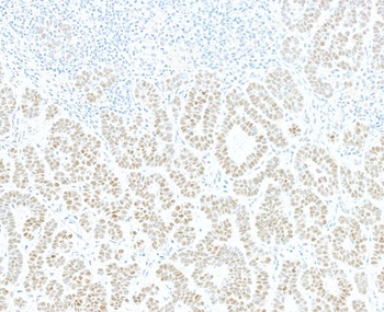
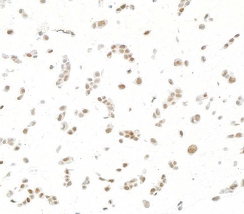
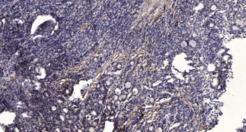
Detection of human JDP2 by immunohistochemistry. Sample: FFPE section of human skin carcinoma. Antibody: Rabbit anti-JDP2 recombinant monoclonal antibody (orb1733245). Secondary: HRP-conjugated goat anti-rabbit IgG.
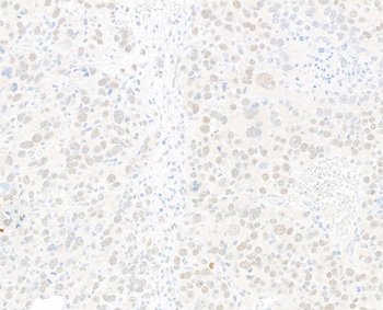
Detection of mouse JDP2 by immunohistochemistry. Sample: FFPE section of mouse RenCa tumor. Antibody: Rabbit anti-JDP2 recombinant monoclonal antibody (orb1733245). Secondary: HRP-conjugated goat anti-rabbit IgG.

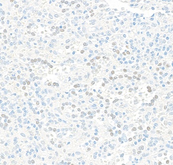
Detection of human JDP2 by immunohistochemistry. Sample: FFPE section of human spleen. Antibody: Rabbit anti-JDP2 recombinant monoclonal antibody (orb1733245). Secondary: HRP-conjugated goat anti-rabbit IgG.

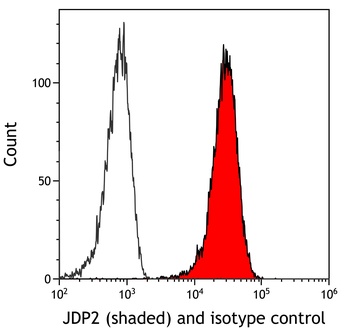
Detection of mouse JDP2 (shaded) in CTLL-2 cells by flow cytometry. Antibody: Rabbit anti-JDP2 recombinant monoclonal antibody (orb1733245) or isotype control (unshaded). Secondary: DyLight® 650-conjugated goat anti-rabbit IgG.

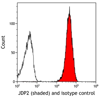
Detection of human JDP2 (shaded) in FaDu cells by flow cytometry. Antibody: Rabbit anti-JDP2 recombinant monoclonal antibody (orb1733245) or isotype control (unshaded). Secondary: DyLight® 650-conjugated goat anti-rabbit IgG.

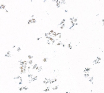
Detection of human JDP2 by immunocytochemistry. Sample: FFPE section of human FaDu cells. Antibody: Rabbit anti-JDP2 recombinant monoclonal antibody (orb1733245). Secondary: HRP-conjugated goat anti-rabbit IgG.
Detection of mouse JDP2 by immunocytochemistry. Sample: FFPE section of mouse C2C12 cells. Antibody: Rabbit anti-JDP2 recombinant monoclonal antibody (orb1733245). Secondary: HRP-conjugated goat anti-rabbit IgG.
- Item 1 of 9
JDP2 Antibody [orb1733243]
FC, ICC, IHC, WB
Human, Mouse
Rabbit
Recombinant
Unconjugated
10 μl (5+ tests) - Item 1 of 3
- Item 1 of 1
- Item 1 of 1

JDP2 (Phospho-T148) antibody [orb570893]
ELISA, IHC
Human, Mouse, Rat
Rabbit
Polyclonal
Unconjugated
50 μg, 100 μg


Submit a review
Filter by Rating

- 5 stars

- 4 stars
- 3 stars
- 2 stars
- 1 stars