You have no items in your shopping cart.
Cart summary
Item 1 of 8
Item 1 of 8
IRAK1 Antibody
Catalog Number: orb1239517
| Catalog Number | orb1239517 |
|---|---|
| Category | Antibodies |
| Description | IRAK1 Antibody |
| Species/Host | Rabbit |
| Clonality | Polyclonal |
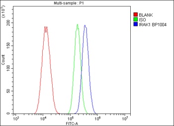
| Tested applications | ELISA, ICC, IF, IP, WB |
| Predicted Reactivity | Bovine |
| Reactivity | Human, Mouse, Rat |
| Isotype | IgG |
| Immunogen | Anti-IRAK antibody (orb1239517) was raised against a peptide corresponding to 13 amino acids near the carboxy terminus of human IRAK. The immunogen is located within the last 50 amino acids of IRAK. |
| Concentration | 1 mg/mL |
| Dilution range | WB: 1-4 μg/mL; IF: 20 μg/mL; ICC: 10 μg/mL.Antibody validated: Western Blot in human, mouse and rat samples; Immunofluorescence and Immunocytochemistry in human samples. All other applications and species not yet tested. |
| Form/Appearance | Liquid |
| Conjugation | Unconjugated |
| MW | Predicted: 77kDObserved: 77 kD |
| Target | IRAK1 |
| UniProt ID | P51617 |
| NCBI | P51617 |
| Storage | IRAK antibody can be stored at 4°C for three months and -20°C, stable for up to one year. As with all antibodies care should be taken to avoid repeated freeze thaw cycles. Antibodies should not be exposed to prolonged high temperatures. |
| Buffer/Preservatives | IRAK Antibody is supplied in PBS containing 0.02% sodium azide. |
| Alternative names | IRAK Antibody: IRAK, pelle, IRAK, Interleukin-1 re Read more... |
| Note | For research use only |
| Expiration Date | 12 months from date of receipt. |
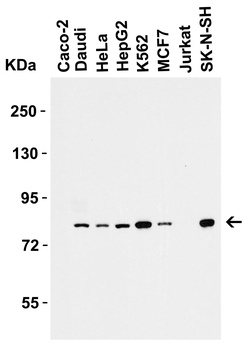
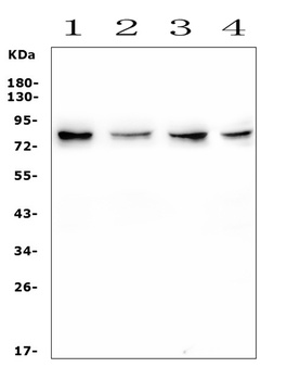
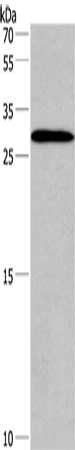
Western Blot Validation in Human Cell Lines. Loading: 15 µg of lysates per lane. Antibodies: IRAK orb1239517 (1 µg/mL), 1h incubation at RT in 5% NFDM/TBST. Secondary: Goat anti-rabbit IgG HRP conjugate at 1:10000 dilution.
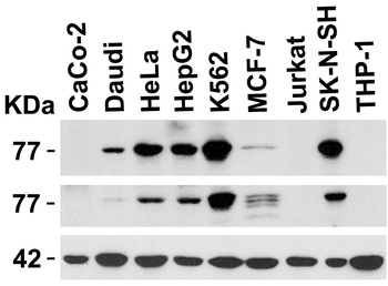
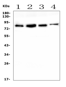
Independent Antibody Validation (IAV) via Protein Expression Profile in Cell Lines. Loading: 15 µg of lysates per lane. Antibodies: IRAK orb1239517 (1 µg/mL), IRAK orb1271054 (2 µg/mL), beta-actin (1 µg/mL), 1h incubation at RT in 5% NFDM/TBST. Secondary: Goat anti-rabbit IgG HRP conjugate at 1:10000 dilution.
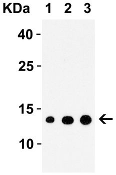
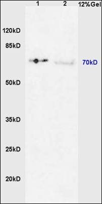
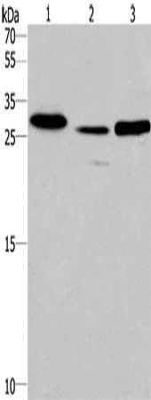
Western Blot Validation with Recombinant Protein. Loading: 30 ng of human IRAK recombinant protein per lane. Antibodies: IRAK orb1239517 (1: 1 µg/mL, 2: 2 µg/mL and 3: 4 µg/mL), 1h incubation at RT in 5% NFDM/TBST. Secondary: Goat anti-rabbit IgG HRP conjugate at 1:10000 dilution.
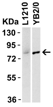
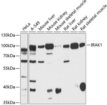
Species Activity in Mouse and Rat Cell Lines. Loading: 15 µg of lysates per lane. Antibodies: IRAK orb1239517 (1 µg/mL, ), 1h incubation at RT in 5% NFDM/TBST. Secondary: Goat anti-rabbit IgG HRP conjugate at 1:10000 dilution.
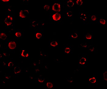
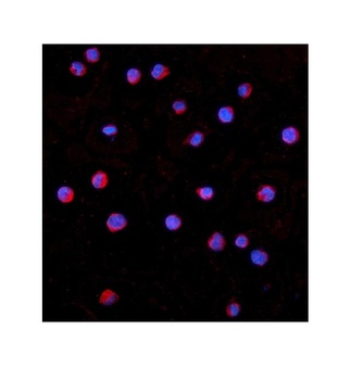
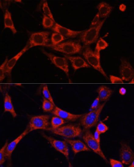
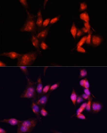
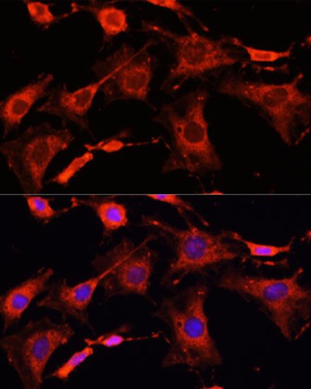
Immunofluorescence Validation of IRAK in Human HeLa Cells. Immunofluorescent analysis of 4% paraformaldehyde-fixed HeLa Cells labeling IRAK with orb1239517 at 20 µg/mL, followed by goat anti-rabbit IgG secondary antibody at 1/500 dilution (red).
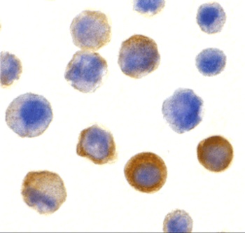
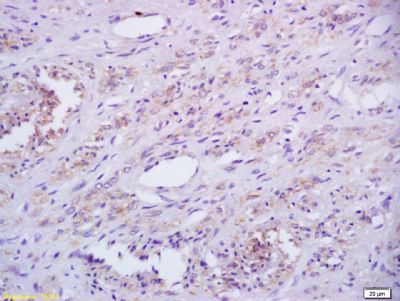
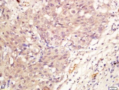
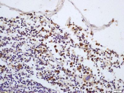
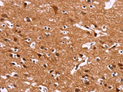
Immunocytochemistry Validation of IRAK in Human HeLa Cells. Immunocytochemical analysis of HeLa cells using anti-IRAK antibody (orb1239517) at 10 µg/ml. Cells was fixed with formaldehyde and blocked with 10% serum for 1 h at RT; antigen retrieval was by heat mediation with a citrate buffer (pH6). Samples were incubated with primary antibody overnight at 4°C. A goat anti-rabbit IgG H&L (HRP) at 1/250 was used as secondary. Counter stained with Hematoxylin.
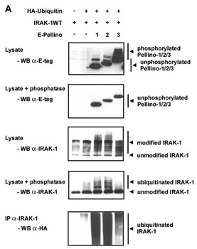
Immunoprecipitation and Overexpression Validation in HEK293T Cells (Schauvliege et al., 2006). Co-expression of Pellino proteins and IRAK-1 leads to Pellino phosphorylation and IRAK-1 polyubiquitination. (A) E-tagged Pellino proteins were co-expressed with IRAK-1WT and HA–ubiquitin in HEK293T cells. For assessment of IRAK-1 polyubiquitination, the same cellextracts, untreated or treated with phosphatase as described above, were analysed for slower migrating forms of IRAK-1 by Western blotting withanti-IRAK-1 (orb1239517). Ubiquitination was specifically detected by IRAK-1 immunoprecipitation followed by Western blotting with anti-HA antibodies.
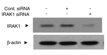
KD Validation in Human Chondrocytes (Ahmad et al., 2007). Chondrocytes were transfected with 250 nM of IRAK1 or control siRNA for 48 h and lysates were analyzed for IRAK1 or β-actin expression levels by immunoblotting. IRAK1 signal was disrupted in IRAK1 KD lysate.
- Item 1 of 6
IRAK1 (phospho-Thr387) antibody [orb6223]
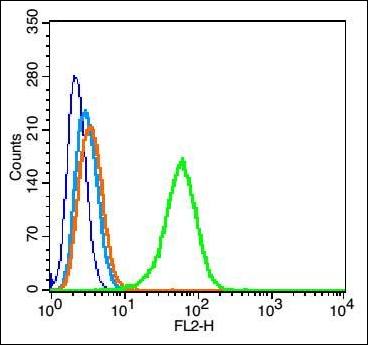
FC, IHC-P, WB
Bovine, Canine, Porcine, Rabbit
200 μl, 50 μl, 100 μl - Item 1 of 6
IRAK-1/IRAK1 Antibody [orb570392]
FC, ICC, IF, IHC, WB
Human, Mouse, Rat
Rabbit
Polyclonal
Unconjugated
10 μg, 100 μg - Item 1 of 3
- Item 1 of 3
- Item 1 of 4
Submit a review
Filter by Rating

- 5 stars

- 4 stars
- 3 stars
- 2 stars
- 1 stars