You have no items in your shopping cart.
NCAM / CD56 Antibody
SKU: orb534487
Description
Images & Validation
−Item 1 of 5
| Tested Applications | IF, IHC-P, WB |
|---|---|
| Dilution range | Immunofluorescence: 1-2ug/ml,Immunohistochemistry (FFPE): 1-2ug/ml for 30 min at RT,Western blot: 1-2ug/ml |
| Reactivity | Human |
| Application Notes |
Key Properties
−| Antibody Type | Primary Antibody |
|---|---|
| Host | Mouse |
| Clonality | Monoclonal |
| Isotype | Mouse IgG1, kappa |
| Clone No. | 56C04, also called 123A8 |
| Immunogen | A membrane preparation of small cell lung carcinoma was used as the immunogen for this CD56 antibody. This mAb reacts with an extracellular domain (close to transmembrane) of CD56/NCAM. |
| Conjugation | Unconjugated |
Storage & Handling
−| Storage | Maintain refrigerated at 2-8°C for up to 2 weeks. For long term storage store at -20°C in small aliquots to prevent freeze-thaw cycles. |
|---|---|
| Disclaimer | For research use only |
Similar Products
−- Item 1 of 1

Mouse NCAM / CD56 Antibody [orb98391]
ICC, IHC-Fr, IHC-P, WB
Human, Zebrafish
Mouse
Monoclonal
Unconjugated
0.1 mg- Item 1 of 4
- Item 1 of 2


Quality Guarantee
Explore bioreagents carefree to elevate your research. All our products are rigorously tested for performance. If a product does not perform as described on its datasheet, our scientific support team will provide expert troubleshooting, a prompt replacement, or a refund. For full details, please see our Terms & Conditions and Buying Guide. Contact us at [email protected].

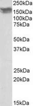
Western blot testing of human brain tissue with CD56 antibody (clone 56C04/123A8). Predicted molecular weight: ~110 kDa (soluble fragment), ~120/125 kDa (GPI-anchored), 140/180 kDa (transmembrane isoforms).
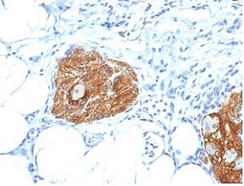
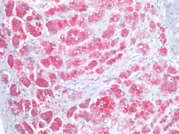
IHC staining of FFPE human pancreas tissue with CD56 antibody (clone 56C04/123A8). HIER: boil tissue sections in pH9 EDTA buffer for 10-20 min followed by cooling at RT for 20 min.
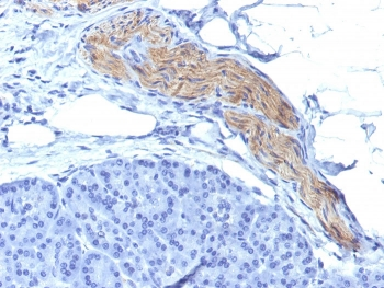
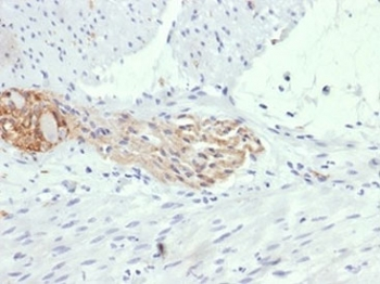
IHC staining of FFPE human thyroid tissue with CD56 antibody (clone 56C04/123A8). HIER: boil tissue sections in pH9 EDTA buffer for 10-20 min followed by cooling at RT for 20 min.
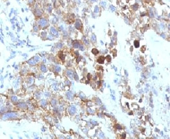
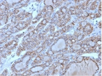
IHC staining of FFPE human colon tissue with CD56 antibody (clone 56C04/123A8). HIER: boil tissue sections in pH9 EDTA buffer for 10-20 min followed by cooling at RT for 20 min.
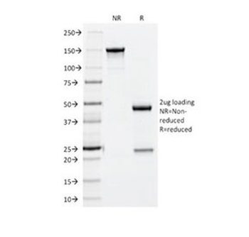
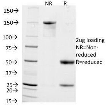
SDS-PAGE analysis of purified, BSA-free CD56 antibody (clone 56C04/123A8) as confirmation of integrity and purity.
Quick Database Links
UniProt
UniProt Details
− No UniProt data available
Documents Download
Datasheet
Product Information
Request a Document
Protocol Information
WB
Western Blot (IB, immunoblot)
IHC-P
Immunohistochemistry Paraffin
IF
Immunofluorescence
NCAM / CD56 Antibody (orb534487)

- 0.0
Based on 0 reviews
Participating in our Biorbyt product reviews program enables you to support fellow scientists by sharing your firsthand experience with our products.
Login to Submit a ReviewAvailable Sizes
Select a size below