You have no items in your shopping cart.
IDE Antibody (Center)
SKU: orb1935700
Description
Research Area
Cardiovascular Research, Cell Biology, Infectious Disease & Virology, Metabolism Research, Neuroscience, Signal Transduction
Images & Validation
−Item 1 of 5
| Tested Applications | IHC-P, WB |
|---|---|
| Dilution Range | WB - 1:1000, IHC-P - 1:100-500 |
| Reactivity | Human, Mouse, Rat |
| Predicted Reactivity | Bovine |
Key Properties
−| Antibody Type | Primary Antibody |
|---|---|
| Host | Rabbit |
| Clonality | Polyclonal |
| Isotype | Rabbit IgG |
| Molecular Weight | 117968 Da |
| Conjugation | Unconjugated |
Storage & Handling
−| Storage | Maintain refrigerated at 2-8°C for up to 2 weeks. For long term storage store at -20°C in small aliquots to prevent freeze-thaw cycles |
|---|---|
| Form/Appearance | Purified polyclonal antibody supplied in PBS with 0.09% (W/V) sodium azide. This antibody is prepared by Saturated Ammonium Sulfate (SAS) precipitation followed by dialysis against PBS. |
| Disclaimer | For research use only |
Quality Guarantee
Explore bioreagents carefree to elevate your research. All our products are rigorously tested for performance. If a product does not perform as described on its datasheet, our scientific support team will provide expert troubleshooting, a prompt replacement, or a refund. For full details, please see our Terms & Conditions and Buying Guide. Contact us at [email protected].
Western blot analysis of anti-IDE Antibody (Center) in A375 cell line lysates (35 ug/lane). IDE (arrow) was detected using the purified Pab.

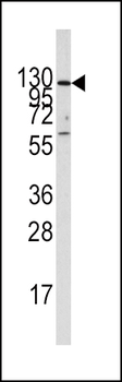
Western blot analysis of lysate from K562 cell line, using IDE Antibody (Center). Diluted at 1:1000. A goat anti-rabbit IgG H&L (HRP) at 1:5000 dilution was used as the secondary antibody. Lysate at 35ug.
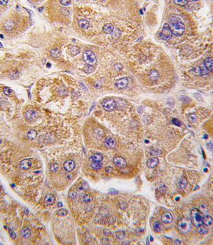
Formalin-fixed and paraffin-embedded human hepatocarcinoma tissue reacted with IDE antibody (Center), which was peroxidase-conjugated to the secondary antibody, followed by DAB staining. This data demonstrates the use of this antibody for immunohistochemistry; clinical relevance has not been evaluated.
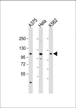
All lanes: Anti-IDE Antibody (Center) at 1:2000 dilution. Lane 1: A375 whole cell lysate. Lane 2: Hela whole cell lysate. Lane 3: K562 whole cell lysate. Lysates/proteins at 20 µg per lane. Secondary Goat Anti-Rabbit IgG, (H+L), Peroxidase conjugated at 1/10000 dilution. Predicted band size: 118 kDa. Blocking/Dilution buffer: 5% NFDM/TBST.
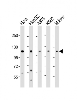
All lanes: Anti-IDE Antibody (Center) at 1:2000 dilution. Lane 1: Hela whole cell lysate. Lane 2: HepG2 whole cell lysate. Lane 3: A375 whole cell lysate. Lane 4: K562 whole cell lysate. Lane 5: mouse liver lysate. Lysates/proteins at 20 µg per lane. Secondary Goat Anti-Rabbit IgG, (H+L), Peroxidase conjugated at 1/10000 dilution. Predicted band size: 118 kDa. Blocking/Dilution buffer: 5% NFDM/TBST.
Quick Database Links
UniProt
UniProt Details
− No UniProt data available
Documents Download
Datasheet
Product Information
Request a Document
Protocol Information
WB
Western Blot (IB, immunoblot)
IHC-P
Immunohistochemistry Paraffin
IDE Antibody (Center) (orb1935700)

- 0.0
Based on 0 reviews
Participating in our Biorbyt product reviews program enables you to support fellow scientists by sharing your firsthand experience with our products.
Login to Submit a ReviewAvailable Sizes
Select a size below