You have no items in your shopping cart.
CD137 Antibody
SKU: orb2650307
Description
Images & Validation
−Item 1 of 2
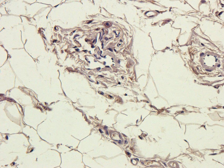
| Tested Applications | IF, WB |
|---|---|
| Dilution range | WB: WB (1/500 - 1/1000), IF/IC (1/100 - 1/500), IF: WB (1/500 - 1/1000), IF/IC (1/100 - 1/500) |
| Reactivity | Human, Mouse, Rat |
Key Properties
−| Host | Rabbit |
|---|---|
| Clonality | Polyclonal |
| Clone No. | TNFRSF9 |
| Conjugation | Unconjugated |
Storage & Handling
−| Storage | Maintain refrigerated at 2-8°C for up to 2 weeks. For long term storage store at -20°C in small aliquots to prevent freeze-thaw cycles. |
|---|---|
| Disclaimer | For research use only |
Alternative Names
−CD137; ILA; Tumor necrosis factor receptor superfamily member 9; 4-1BB ligand receptor; CDw137; T-cell antigen 4-1BB homolog; T-cell antigen ILA; CD137
Similar Products
−- Item 1 of 8
CD137 Antibody / 4-1BB / TNFRSF9 [orb607142]
ELISA, FACS, IF, IHC-P, WB
Human
Mouse
Monoclonal
Unconjugated
100 μg, 20 μg - Item 1 of 8
CD137 Antibody / 4-1BB / TNFRSF9 [orb2642859]
ELISA, FACS, IF, IHC-P, WB
Human
Mouse
Monoclonal
Unconjugated
100 μg - Item 1 of 6
CD137 antibody [orb389316]
ICC, IF, IHC-P, WB
Guinea pig, Mouse, Rat
Rabbit
Polyclonal
Unconjugated
100 μg - Item 1 of 1
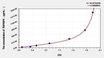
Mouse Tumor Necrosis Factor Receptor Superfamily, Member 9 (TNFRSF9) ELISA Kit [orb782213]
Mouse
23.5-1500 pg/mL
6 pg/mL
48 T, 96 T - Item 1 of 1
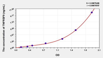
Human Tumor Necrosis Factor Receptor Superfamily, Member 9 (TNFRSF9) ELISA Kit [orb779046]
Human
0.16-10 ng/mL
0.062 ng/mL
96 T, 48 T


Quality Guarantee
Explore bioreagents carefree to elevate your research. All our products are rigorously tested for performance. If a product does not perform as described on its datasheet, our scientific support team will provide expert troubleshooting, a prompt replacement, or a refund. For full details, please see our Terms & Conditions and Buying Guide. Contact us at [email protected].
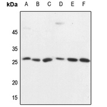
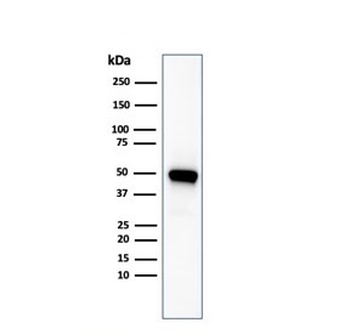
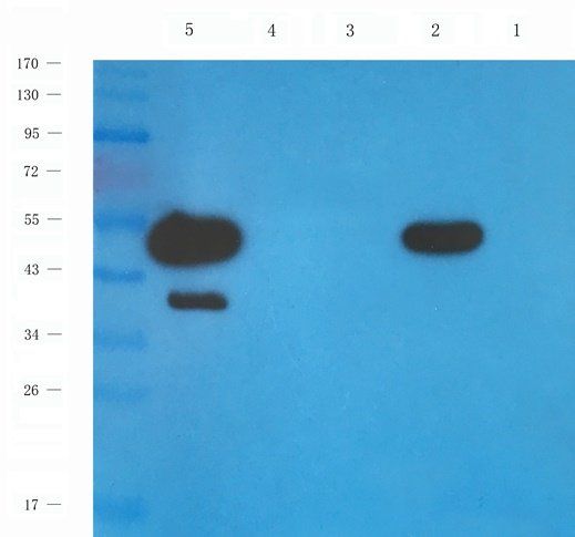
Western blot analysis of CD137 expression in HEK293T (A), Hela (B), HepG2 (C), mouse brain (D), mouse spleen (E), rat spleen (F) whole cell lysates.
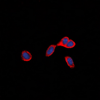
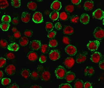
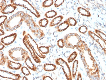
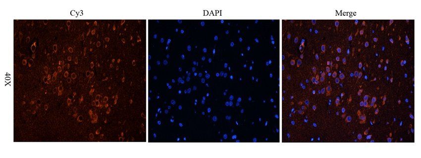
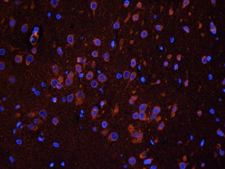
Immunofluorescent analysis of CD137 staining in HepG2 cells. Formalin-fixed cells were permeabilized with 0.1% Triton X-100 in TBS for 5-10 minutes and blocked with 3% BSA-PBS for 30 minutes at room temperature. Cells were probed with the primary antibody in 3% BSA-PBS and incubated overnight at 4°C in a hidified chamber. Cells were washed with PBST and incubated with a DyLight 594-conjugated secondary antibody (red) in PBS at room temperature in the dark. DAPI was used to stain the cell nuclei (blue).
Quick Database Links
UniProt
UniProt Details
− No UniProt data available
Documents Download
Datasheet
Product Information
Request a Document
Protocol Information
WB
Western Blot (IB, immunoblot)
IF
Immunofluorescence
CD137 Antibody (orb2650307)

- 0.0
Based on 0 reviews
Participating in our Biorbyt product reviews program enables you to support fellow scientists by sharing your firsthand experience with our products.
Login to Submit a ReviewAvailable Sizes
Select a size below