You have no items in your shopping cart.
Cart summary
Item 1 of 4
Item 1 of 4
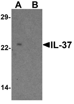
IL37 Antibody
Catalog Number: orb738386
| Catalog Number | orb738386 |
|---|---|
| Category | Antibodies |
| Description | IL37 Antibody |
| Species/Host | Rabbit |
| Clonality | Polyclonal |
| Tested applications | ELISA, FC, ICC, IF, IHC |
| Reactivity | Human |
| Isotype | Rabbit IgG |
| Immunogen | E.coli-derived human IL37 recombinant protein (Position: V46-D218). |
| Concentration | Adding 0.2 ml of distilled water will yield a concentration of 500 μg/ml. |
| Dilution range | Immunohistochemistry (Paraffin-embedded Section), 2-5μg/ml, Human Immunocytochemistry/Immunofluorescence, 5μg/ml, Human Flow Cytometry, 1-3μg/1x106 cells, Human Direct ELISA, 0.1-0.5μg/ml, Human |
| Form/Appearance | Lyophilized |
| Conjugation | Unconjugated |
| UniProt ID | Q9NZH6 |
| Storage | Store at -20˚C for one year from date of receipt. After reconstitution, at 4˚C for one month. It can also be aliquotted and stored frozen at -20˚C for six months. Avoid repeated freeze-thaw cycles. |
| Note | For research use only |
| Application notes | Tested Species: In-house tested species with positive results. Other applications have not been tested. Optimal dilutions should be determined by end users. Add 0.2ml of distilled water will yield a concentration of 500ug/ml. |
| Expiration Date | 12 months from date of receipt. |
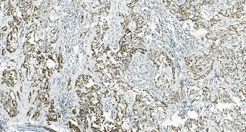
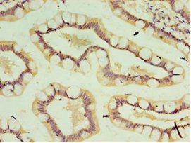
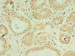
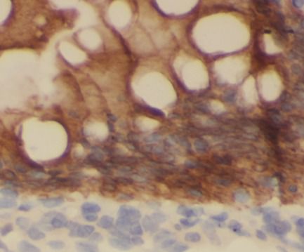
IHC analysis of IL37 using anti-IL37 antibody (orb738386). IL37 was detected in paraffin-embedded section of human B lymphocytic tumor tissue. Heat mediated antigen retrieval was performed in EDTA buffer (pH8.0, epitope retrieval solution). The tissue section was blocked with 10% goat serum. The tissue section was then incubated with 2μg/ml rabbit anti-IL37 Antibody (orb738386) overnight at 4°C. Biotinylated goat anti-rabbit IgG was used as secondary antibody and incubated for 30 minutes at 37°C. The tissue section was developed using Strepavidin-Biotin-Complex (SABC) (Catalog # orb90444) with DAB as the chromogen.
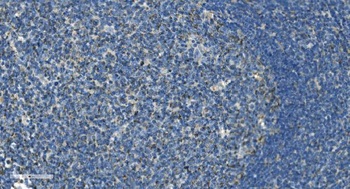
IHC analysis of IL37 using anti-IL37 antibody (orb738386). IL37 was detected in paraffin-embedded section of human tonsil tissue. Heat mediated antigen retrieval was performed in EDTA buffer (pH8.0, epitope retrieval solution). The tissue section was blocked with 10% goat serum. The tissue section was then incubated with 2μg/ml rabbit anti-IL37 Antibody (orb738386) overnight at 4°C. Biotinylated goat anti-rabbit IgG was used as secondary antibody and incubated for 30 minutes at 37°C. The tissue section was developed using Strepavidin-Biotin-Complex (SABC) (Catalog # orb90444) with DAB as the chromogen.
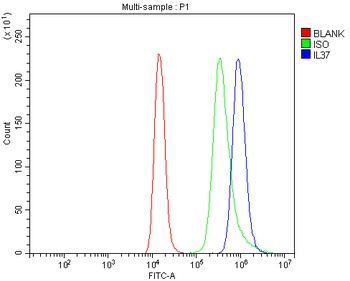
Flow Cytometry analysis of Raji cells using anti-IL37 antibody (orb738386). Overlay histogram showing Raji cells stained with orb738386 (Blue line). The cells were blocked with 10% normal goat serum. And then incubated with rabbit anti-IL37 Antibody (orb738386, 1μg/1x10^6 cells) for 30 min at 20°C. DyLight®488 conjugated goat anti-rabbit IgG (5-10μg/1x10^6 cells) was used as secondary antibody for 30 minutes at 20°C. Isotype control antibody (Green line) was rabbit IgG (1μg/1x10^6) used under the same conditions. Unlabelled sample (Red line) was also used as a control.
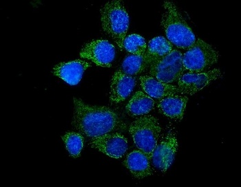
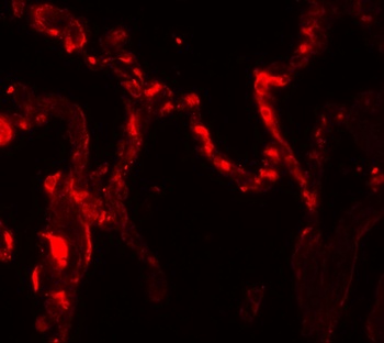
IF analysis of IL37 using anti-IL37 antibody (orb738386). IL37 was detected in immunocytochemical section of A431 cells. Enzyme antigen retrieval was performed using IHC enzyme antigen retrieval reagent (orb90553) for 15 mins. The cells were blocked with 10% goat serum. And then incubated with 5μg/mL rabbit anti-IL37 Antibody (orb738386) overnight at 4°C. DyLight®488 Conjugated Goat Anti-Rabbit IgG was used as secondary antibody at 1:100 dilution and incubated for 30 minutes at 37°C. The section was counterstained with DAPI. Visualize using a fluorescence microscope and filter sets appropriate for the label used.
- Item 1 of 2
- Item 1 of 3
- Item 1 of 1
- Item 1 of 2

Submit a review
Filter by Rating

- 5 stars

- 4 stars
- 3 stars
- 2 stars
- 1 stars