You have no items in your shopping cart.
Cart summary
Item 1 of 8
Item 1 of 8
IgM Antibody
Catalog Number: orb1806431
| Catalog Number | orb1806431 |
|---|---|
| Category | Antibodies |
| Description | IgM Antibody |
| Species/Host | Rabbit |
| Clonality | Recombinant |
| Tested applications | FC, ICC, IHC, IP, WB |
| Reactivity | Human, Mouse |
| Immunogen | Human IgM |
| Concentration | 100 µg/ml |
| Dilution range | WB: 1:1000, IP: 20 µl/mg lysate, IHC: 1:100 to 1:500. Epitope retrieval with citrate buffer pH 6.0 is recommended for FFPE tissue sections, ICC: 1:100 to 1:500. Epitope retrieval with citrate buffer pH 6.0 is recommended for FFPE cell sections |
| Form/Appearance | Whole IgG |
| Conjugation | Unconjugated |
| Target | IgM |
| Storage | Shelf life: 1 year from date of receipt. Storage: 2 - 8°C |
| Buffer/Preservatives | Borate Buffered Saline (BBS) pH 8.2 with 0.1% rAlbumin and 0.09% Sodium Azide |
| Note | For research use only |
| Application notes | Format: Whole IgG |
| Expiration Date | 12 months from date of receipt. |

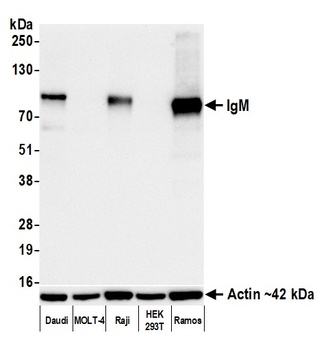
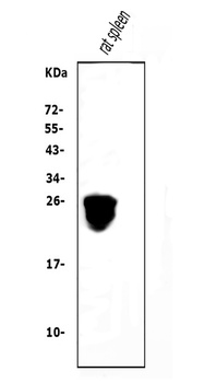
Detection of human IgM by western blot. Samples: Whole cell lysate (10 µg) from Daudi, MOLT-4, Raji, HEK293T, and Ramos cells prepared using NETN lysis buffer. Antibody: Rabbit anti-IgM recombinant monoclonal antibody (orb1806431) used at 1:1000. Secondary: HRP-conjugated goat anti-rabbit IgG. Detection: Chemiluminescence with an exposure time of 10 seconds. Lower Panel: Rabbit anti-Actin recombinant monoclonal antibody.

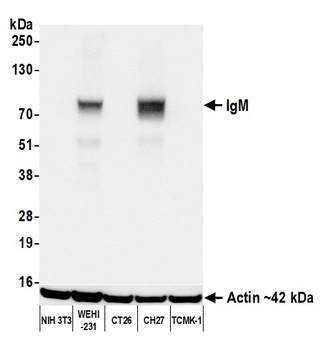
Detection of mouse IgM by western blot. Samples: Whole cell lysate (50 µg) from NIH 3T3, WEHI-231, CT26, CH27, and TCMK-1 cells prepared using NETN lysis buffer. Antibody: Rabbit anti-IgM recombinant monoclonal antibody (orb1806431) used at 1:1000. Secondary: HRP-conjugated goat anti-rabbit IgG. Detection: Chemiluminescence with an exposure time of 1 second. Lower Panel: Rabbit anti-Actin recombinant monoclonal antibody.
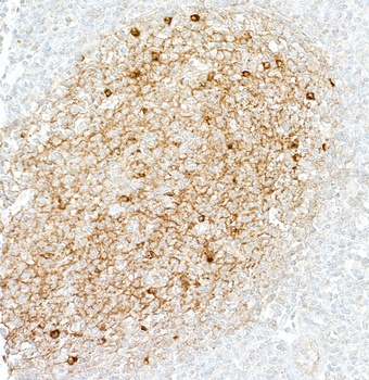
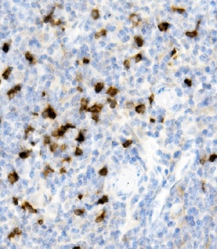
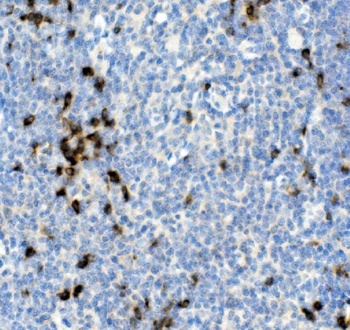
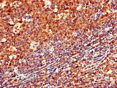
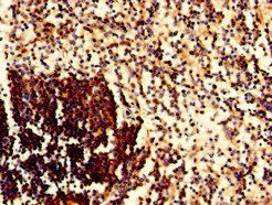
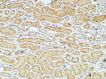
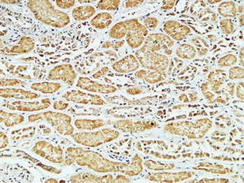
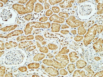
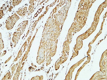
Detection of human IgM by immunohistochemistry. Sample: FFPE section of tonsil. Antibody: Rabbit anti-IgM recombinant monoclonal antibody (orb1806431). Secondary: HRP-conjugated goat anti-rabbit IgG.
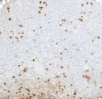
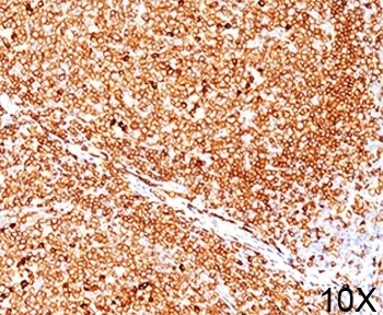
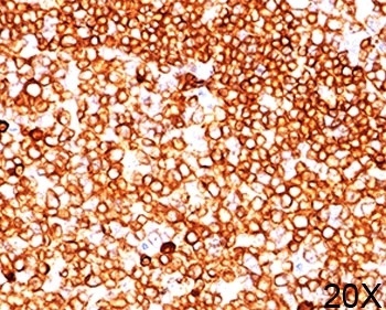
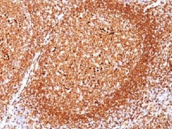
Detection of human IgM by immunohistochemistry. Sample: FFPE section of B-cell lymphoma. Antibody: Rabbit anti-IgM recombinant monoclonal antibody (orb1806431). Secondary: HRP-conjugated goat anti-rabbit IgG.

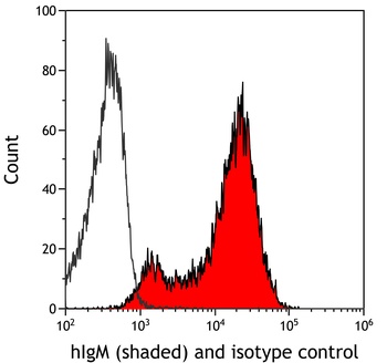
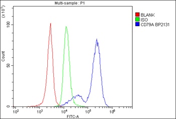
Detection of human IgM (shaded) in Raji cells by flow cytometry. Antibody: Rabbit anti-IgM recombinant monoclonal antibody (orb1806431) or isotype control (unshaded). Secondary: DyLight® 650-conjugated goat anti-rabbit IgG.

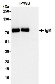
Detection of human IgM by western blot of immunoprecipitates. Samples: Whole cell lysate (1.0 mg per IP reaction; 20% of IP loaded) from Ramos cells prepared using NETN lysis buffer. Antibodies: Rabbit anti-IgM recombinant monoclonal antibody (orb1806431) used for IP at 20 µl/mg lysate.

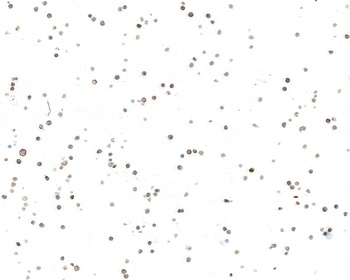
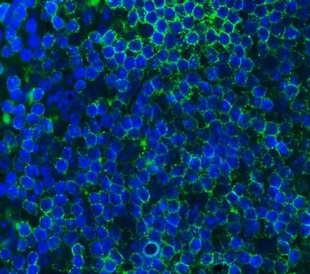
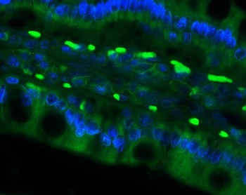
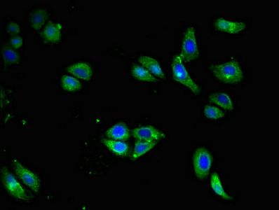
Detection of human IgM by immunocytochemistry. Sample: FFPE section of Daudi cells. Antibody: Rabbit anti-IgM recombinant monoclonal antibody (orb1806431). Secondary: HRP-conjugated goat anti-rabbit IgG.

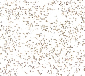
Detection of mouse IgM by immunocytochemistry. Sample: FFPE section of WEHI-231 cells. Antibody: Rabbit anti-IgM recombinant monoclonal antibody (orb1806431). Secondary: HRP-conjugated goat anti-rabbit IgG.
- Item 1 of 8
- Item 1 of 6
Cd79a Antibody [orb654317]
ELISA, FC, IF, IHC, WB
Mouse, Rat
Rabbit
Polyclonal
Unconjugated
10 μg, 100 μg - Item 1 of 3
- Item 1 of 4
- Item 1 of 4


Submit a review
Filter by Rating

- 5 stars

- 4 stars
- 3 stars
- 2 stars
- 1 stars