You have no items in your shopping cart.
Cart summary
Item 1 of 3
Item 1 of 3
IGKV1OR1-1 Antibody
Catalog Number: orb1672202
| Catalog Number | orb1672202 |
|---|---|
| Category | Antibodies |
| Description | IGKV1OR1-1 Antibody |
| Clonality | Recombinant |
| Clone Number | PAb421 |
| Tested applications | ELISA, IF, IHC-Fr, IHC-P, IP, In vivo, WB |
| Reactivity | Human, Monkey, Mouse, Rabbit, Rat |
| Isotype | IgG kappa |
| Immunogen | Synthetic peptide corresponding to aa 371-380 of human p53. |
| Concentration | batch dependent |
| Conjugation | Unconjugated |
| Target | IGKV1OR1-1 |
| UniProt ID | P04637 |
| Storage | Store at 4°C for up to 3 months. For longer storage, aliquot and store at -20°C. |
| Buffer/Preservatives | PBS with 0.02% Proclin 300. |
| Alternative names | Tumor suppressor p53, Cellular tumor antigen p53 Read more... |
| Note | For research use only |
| Expiration Date | 12 months from date of receipt. |
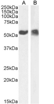
Western Blot using anti-p53 antibody P00142. A431 cell nuclear (A) and cytoplasmic (B) extract (35 ug protein in RIPA buffer) was resolved on a 10% SDS PAGE gel and blots probed with the chimeric rsion of P-180 at 0.1 ug/ml before detection by an anti-rondary antibody. A primary incubation of 1h was used and protein was detected by chemiluminescence. The expected band size for p53 is 43.7 kDa- tho ugh due to the high number of proline residues in this protein runs at a size of ~53kDa (c.f. Ziemer et al.- PMID: 7107651). orb1672202 successfully detected both human nuclear and cytoplasmic p53.
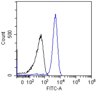
Flow-cytometry using the anti-p53 antibody P00142. Jurkat cells were stained with unimmunized r antibody (black line) or the rmeric version of P-180 - blue line at a concentration of 10 ug/ml for 30 mins at RT. After washing- bound antibody was detected using anti-r JK (FITC-conjugate) antibody (129936) at 2 ug/ml and cells analyzed on a FACSCanto flow-cytometer.
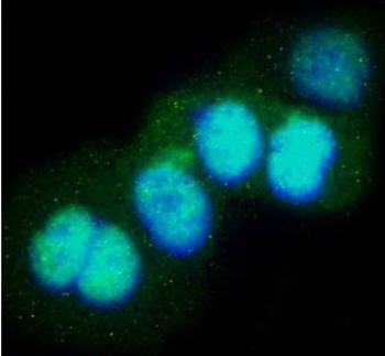
Immunofluorescence staining of fixed A431 with anti-p53 antibody P00142. Immunofluorescence analysis of paraformaldehyde fixed A431 cells- permewith 0.15% Triton and stained with the chimeric r version of P-180 at 10 ug/ml for 1h followed by Alexa Fluor® 488 secondary antibody (1 ug/ml)- showing nuclear staining. The nuclear stain is DAPI (blue). Panels show from left-right- top-bottom orb1672202- DAPI- merged channels and a negative control. The negative control was stained with unimmunized r followed by Alexa Fluor® 488 secondary antibody.

IGKV1OR1-1 Antibody [orb1672203]
ELISA, IF, IHC-Fr, IHC-P, IP, In vivo, WB
Human, Monkey, Mouse, Rabbit, Rat
Recombinant
Unconjugated
0.2 mg
Submit a review
Filter by Rating

- 5 stars

- 4 stars
- 3 stars
- 2 stars
- 1 stars