You have no items in your shopping cart.
Cart summary
Item 1 of 7
Item 1 of 7
IDUA antibody
Catalog Number: orb527236
| Catalog Number | orb527236 |
|---|---|
| Category | Antibodies |
| Description | Rabbit polyclonal antibody to IDUA |
| Species/Host | Rabbit |
| Clonality | Polyclonal |
| Clone Number | RB26860 |
| Tested applications | FC, IHC-P, WB |
| Reactivity | Human |
| Isotype | Rabbit IgG |
| Immunogen | Synthetic Peptide |
| Dilution range | FC: 1:25, WB: 1:1000 |
| Form/Appearance | Purified polyclonal antibody supplied in PBS with 0.09% (W/V) sodium azide. This antibody is purified through a protein A column, followed by peptide affinity purification. |
| Conjugation | Unconjugated |
| MW | 72670 |
| Target | IDUA |
| UniProt ID | P35475 |
| NCBI | NP_000194.2 |
| Alternative names | Alpha-L-iduronidase, IDUA Read more... |
| Note | For research use only |
| Expiration Date | 12 months from date of receipt. |
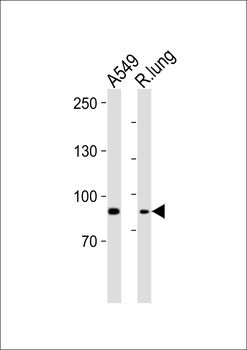
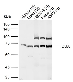
Western blot analysis of lysates from A549 cell line, rat lung tissue lysate (from left to right), using IDUA Antibody (Center) at 1:1000 dilution.
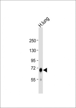
Western blot:Anti-IDUA Antibody (Center) at 1:1000 dilution + human lung lysate.Secondary antibody was Goat Anti-Rabbit IgG, (H+L).
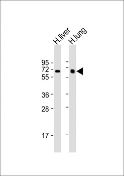
Western blot:All lanes:Anti-IDUA Antibody (Center) at 1:2000 dilution.Lane 1:human liver lysates;Lane 2:human lung lysates.
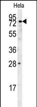
Western blot analysis of Hela cell line lysates using IDUA Antibody (Center).This demonstrates the IDUA antibody detected the IDUA protein (arrow).
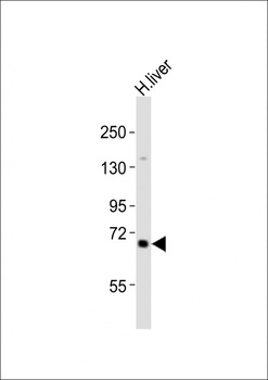
Western blot:Anti-IDUA Antibody (Center) at 1:2000 dilution + human liver lysates.Secondary antibody was Goat Anti-Rabbit IgG, (H+L).
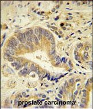
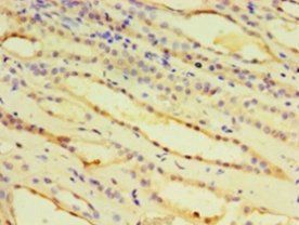
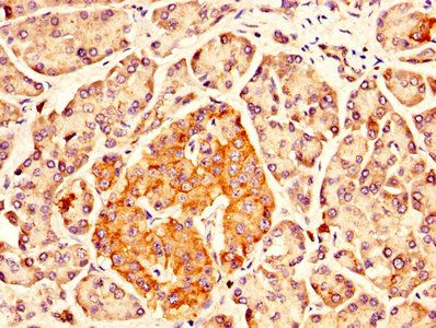
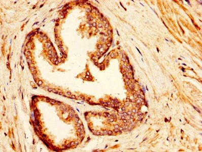
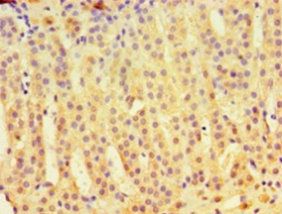
Immunohistochemical analysis of human prostate carcinoma using IDUA Antibody (Center), followed by peroxidase conjugation of the secondary antibody and DAB staining.
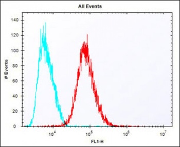
Flow cytometric analysis of HepG2 cells stained with orb527236(red line) at 1:25 dilution.Secondary antibody was Alexa Fluor® 488 goat anti-rabbit lgG (H+L).Isotype control antibody (blue line) was rabbit IgG1.
- Item 1 of 4
- Item 1 of 1

- Item 1 of 1
Submit a review
Filter by Rating

- 5 stars

- 4 stars
- 3 stars
- 2 stars
- 1 stars