You have no items in your shopping cart.
Cart summary
Item 1 of 6
Item 1 of 6
Hsp60/HSPD1 Antibody
Catalog Number: orb251520
| Catalog Number | orb251520 |
|---|---|
| Category | Antibodies |
| Description | Hsp60/HSPD1 Antibody |
| Species/Host | Rabbit |
| Clonality | Polyclonal |
| Tested applications | ICC, IF, IHC, WB |
| Reactivity | Human, Mouse, Rat |
| Isotype | Rabbit IgG |
| Immunogen | E.coli-derived human Hsp60 recombinant protein (Position: A260-Q496). Human Hsp60 shares 97% amino acid (aa) sequence identity with both mouse and rat Hsp60. |
| Concentration | Adding 0.2 ml of distilled water will yield a concentration of 500 μg/ml. |
| Dilution range | Western blot, 0.1-0.5μg/ml, Human, Mouse, Rat Immunohistochemistry (Paraffin-embedded Section), 0.5-1μg/ml, Human, Mouse, Rat, By Heat Immunocytochemistry/Immunofluorescence, 2μg/ml, Human |
| Form/Appearance | Lyophilized |
| Conjugation | Unconjugated |
| MW | 61055 MW |
| UniProt ID | P10809 |
| Storage | Store at -20˚C for one year from date of receipt. After reconstitution, at 4˚C for one month. It can also be aliquotted and stored frozen at -20˚C for six months. Avoid repeated freeze-thaw cycles. |
| Alternative names | 60 kDa heat shock protein, mitochondrial;60 kDa ch Read more... |
| Note | For research use only |
| Application notes | WB: The detection limit for Hsp60 is approximately 0.1ng/lane under reducing conditions. Tested Species: In-house tested species with positive results. By Heat: Boiling the paraffin sections in 10mM citrate buffer, pH6.0, for 20mins is required for the staining of formalin/paraffin sections. Other applications have not been tested. Optimal dilutions should be determined by end users. . Add 0.2ml of distilled water will yield a concentration of 500ug/ml. |
| Expiration Date | 12 months from date of receipt. |
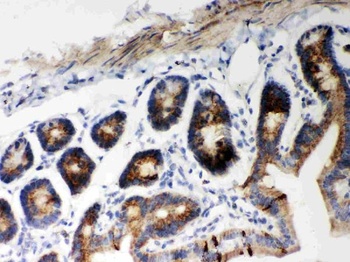
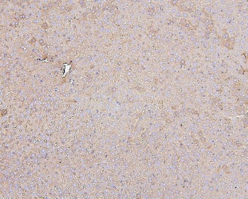
IHC(P) analysis of Mouse Intestine Tissue using Anti-Hsp60 Picoband antibody.
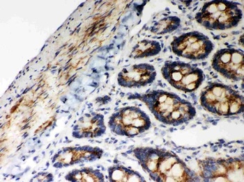
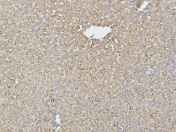
IHC(P) analysis of Rat Intestine Tissue using Anti-Hsp60 Picoband antibody.
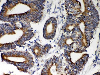
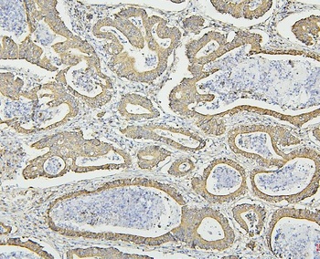
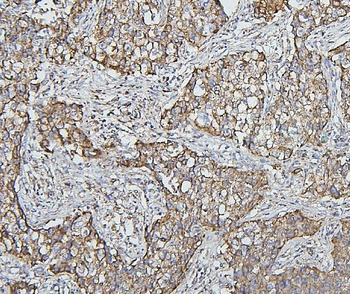
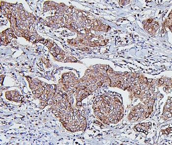
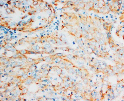
IHC(P) analysis of Human Intestinal Cancer Tissue using Anti-Hsp60 Picoband antibody.
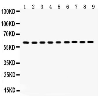
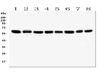
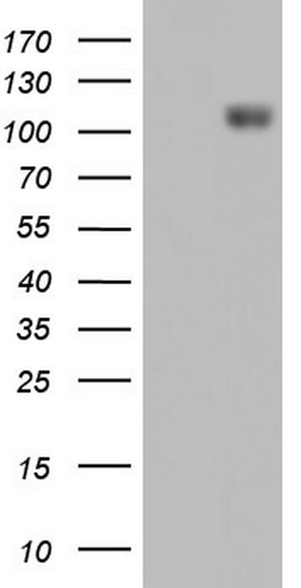
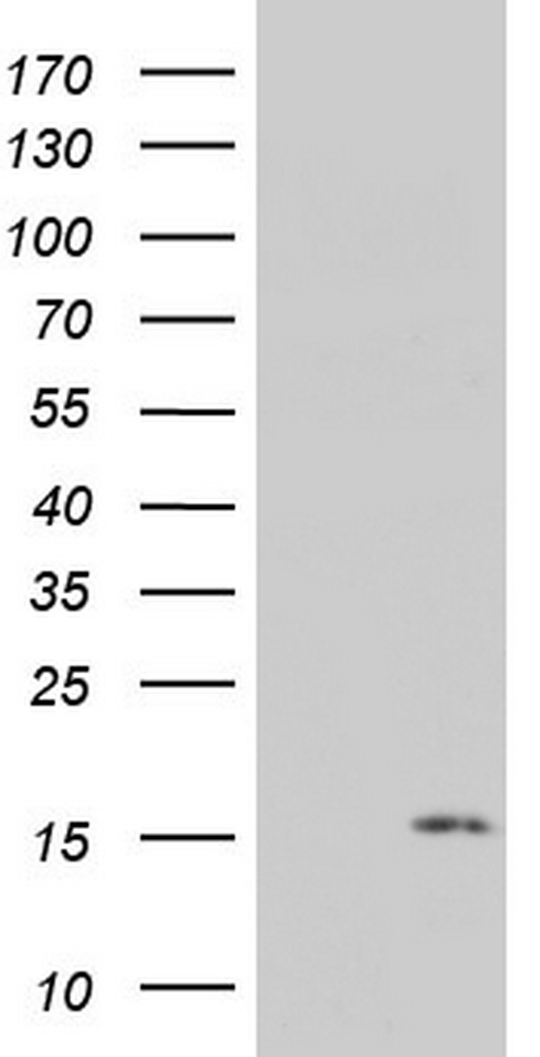
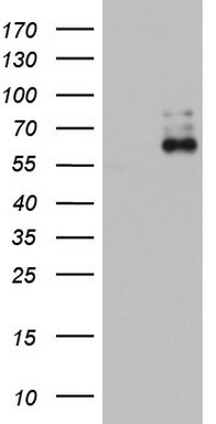
Western blot analysis using Anti-Hsp60 Picoband antibody.Lane 1:Rat Kidney Tissue;2:Mouse Kidney Tissue;3:HEPA Cell;4:NRK Cell;5:PC-12 Cell;6:SW620 Cell;7:A549 Cell;8:A431 Cell;9:HELA Cell.
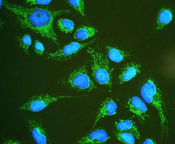
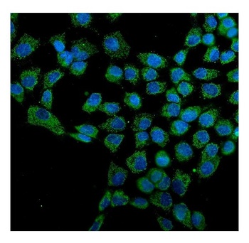
IF analysis of Hsp60 using anti-Hsp60 antibody.Hsp60 was detected in immunocytochemical section of U20S cell.
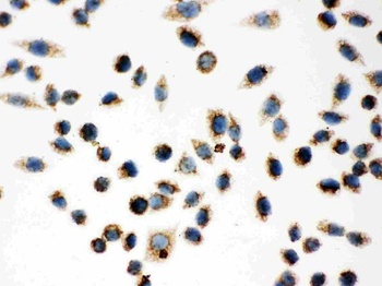
IHC analysis of Hsp60 using anti-Hsp60 antibody.Hsp60 was detected in immunocytochemical section of SMMC-7721 Cell.
- Item 1 of 9
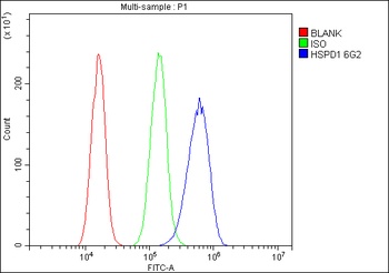
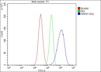
Hsp60/HSPD1 Antibody (monoclonal, 6G2) [orb570314]
FC, ICC, IF, IHC, WB
Human, Mouse, Rat
Mouse
Monoclonal
Unconjugated
10 μg, 100 μg - Item 1 of 2
HSP60/HSPD1 Antibody [orb18084]
FC, ICC, IF, IHC, WB
Equine, Monkey, Rabbit
Human, Mouse, Rat
Rabbit
Polyclonal
Unconjugated
10 μg, 100 μg - Item 1 of 1
- Item 1 of 1
Hsp60 (HSPD1) antibody [orb1319035]
IHC, WB
Canine, Human, Monkey, Mouse, Rat
Mouse
Monoclonal
Unconjugated
100 μl - Item 1 of 1
Hsp60 (HSPD1) antibody [orb1319036]
IHC, WB
Canine, Human, Monkey, Mouse, Rat
Mouse
Monoclonal
Unconjugated
100 μl

Submit a review
Filter by Rating

- 5 stars

- 4 stars
- 3 stars
- 2 stars
- 1 stars