You have no items in your shopping cart.
Cart summary
Item 1 of 3
Item 1 of 3
HSD3B1 Antibody
Catalog Number: orb1672050
| Catalog Number | orb1672050 |
|---|---|
| Category | Antibodies |
| Description | HSD3B1 Antibody |
| Clonality | Recombinant |
| Clone Number | SAIC-34B-124 |
| Tested applications | ELISA, WB |
| Reactivity | Human |
| Isotype | IgG kappa |
| Immunogen | Peptide "LSEDYGVLK", derived from the cellular redox regulator Peroxiredoxin-2, conjugated to KLH. |
| Concentration | batch dependent |
| Conjugation | Unconjugated |
| Target | HSD3B1 |
| UniProt ID | P32119 |
| Storage | Store at 4°C for up to 3 months. For longer storage, aliquot and store at -20°C. |
| Buffer/Preservatives | PBS with 0.02% Proclin 300. |
| Alternative names | Peroxiredoxin-2,, NKEFB, TDPX1 Read more... |
| Note | For research use only |
| Expiration Date | 12 months from date of receipt. |
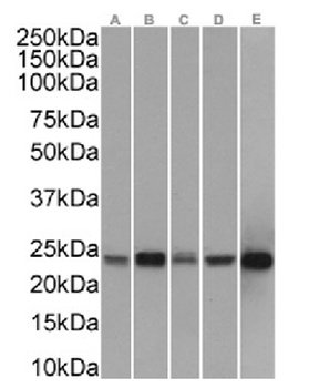
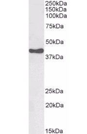
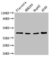
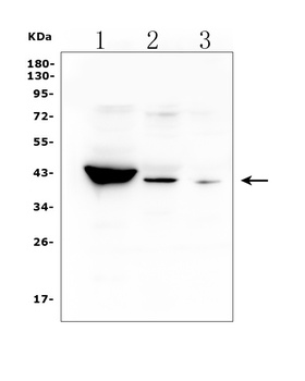
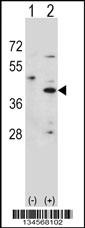
Western Blot using anti-PRDX2 antibody SAIC-34B-124. HepG2(A) (0.003 ug/ml)- HeLa(B) (0.003 ug/ml)- MCF7(C) (0.003 ug/ml)- K562(D) (0.003 ug/ml) and LNCaP (E) (0.001 ug/ml) cell lysates (35 ug protein in RIPA buffer) were resolved on a SDS PAGE gel and blots were probed with the chimeric rsion of SAIC-34B-124 (orb1672050)- before detection using an anti-rondary antibody. A primary incubation of 1h was used and protein was detected by chemiluminescence.
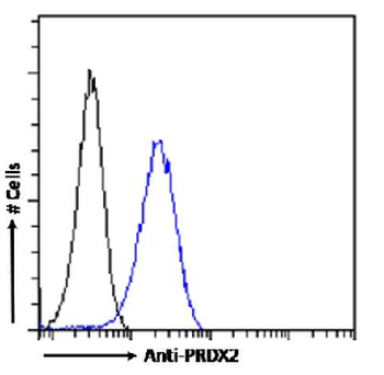
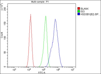
Flow cytometry using the Anti-PRDX2 antibody SAIC-34B-124. Paraformaldehyde fixed HeLa cells permewith 0.5% Triton were stained with anti-unknown specificity antibody (3.0; isotype control - black line) or the r version of SAIC-34B-124 (orb1672050 - blue line) at a dilution of 1:100 for 1h at RT. After washing- the bound antibody was detected using a goat anti-r AlexaFluor® 488 antibody at a dilution of 1:1000 and cells analyzed using a FACSCanto flow-cytometer.
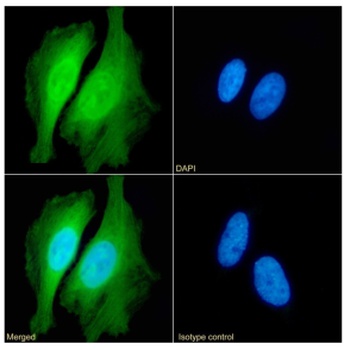
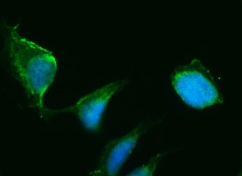
Immunofluorescence staining of HeLa cells with anti-PRDX2 (SAIC-34B-124). Immunofluorescence analysis of paraformaldehyde fixed HeLa cells permewith 0.15% Triton stained with the chimeric r version of SAIC-34B-124 (orb1672050) (1:100 dilution) for 1h followed by Alexa Fluor® 488 secondary antibody (1:1000 dilution)- showing nuclear and cytoplasmic staining. The nuclear stain is DAPI (blue). Panels show from left-right- top-bottom orb1672050- DAPI- merged channels and an isotype control. The isotype control was an unknown specificity antibody (3.0) followed by staining with Alexa Fluor® 488 secondary antibody.
- Item 1 of 4
- Item 1 of 3
- Item 1 of 3
- Item 1 of 4
HSD3B1 Antibody [orb614122]
ELISA, FC, ICC, IF, IHC, WB
Human
Rabbit
Polyclonal
Unconjugated
10 μg, 100 μg - Item 1 of 3

Submit a review
Filter by Rating

- 5 stars

- 4 stars
- 3 stars
- 2 stars
- 1 stars