You have no items in your shopping cart.
Cart summary
Item 1 of 10
Item 1 of 10
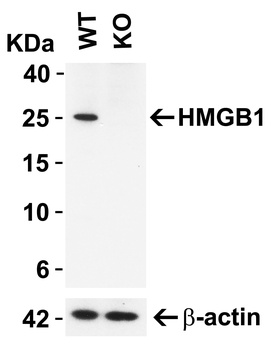
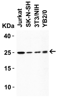
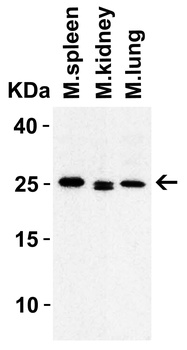
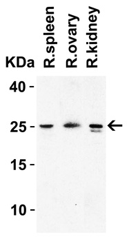
HMGB1 Antibody
Catalog Number: orb389479
| Catalog Number | orb389479 |
|---|---|
| Category | Antibodies |
| Description | HMGB1 Antibody |
| Species/Host | Rabbit |
| Clonality | Polyclonal |
| Tested applications | FC, ICC, IF, IHC, WB |
| Reactivity | Human, Mouse, Rat |
| Isotype | Rabbit IgG |
| Immunogen | A synthetic peptide corresponding to a sequence at the C-terminus of human HMGB1 (124-154aa DVAKKLGEMWNNTAADDKQPYEKKAAKLKEK), identical to the related mouse and rat sequences. |
| Concentration | Adding 0.2 ml of distilled water will yield a concentration of 500 μg/ml. |
| Dilution range | Immunohistochemistry (Paraffin-embedded Section), 0.5-1μg/ml, Human, Mouse, Rat, By Heat Western blot, 0.1-0.5μg/ml, Human, Mouse, RatImmunocytochemistry/Immunofluorescence, 2μg/ml, Human Flow Cytometry, 1-3μg/1x106 cells, Human |
| Form/Appearance | Lyophilized |
| Conjugation | Unconjugated |
| MW | 24894 MW |
| UniProt ID | P09429 |
| Storage | Store at -20˚C for one year from date of receipt. After reconstitution, at 4˚C for one month. It can also be aliquotted and stored frozen at -20˚C for six months. Avoid repeated freeze-thaw cycles. |
| Alternative names | High mobility group protein B1;High mobility group Read more... |
| Note | For research use only |
| Application notes | Tested Species: In-house tested species with positive results. By Heat: Boiling the paraffin sections in 10mM citrate buffer, pH6.0, for 20mins is required for the staining of formalin/paraffin sections. Other applications have not been tested. Optimal dilutions should be determined by end users. Add 0.2ml of distilled water will yield a concentration of 500ug/ml. |
| Expiration Date | 12 months from date of receipt. |
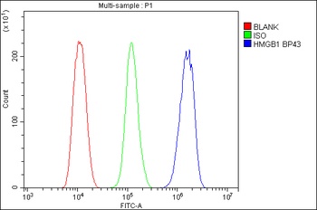
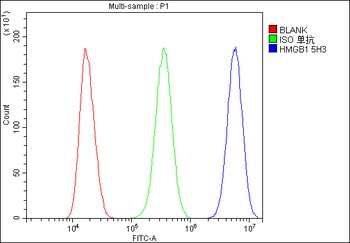
Flow Cytometry analysis of THP-1 cells using anti-HMGB1 antibody (Blue line).Isotype control antibody (Green line) was rabbit IgG .Unlabelled sample (Red line) was also used as a control.
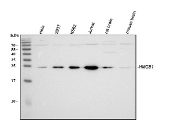
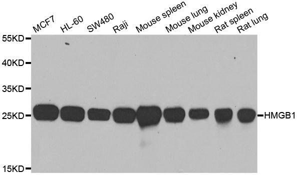
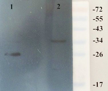
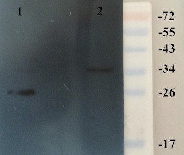
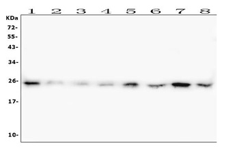
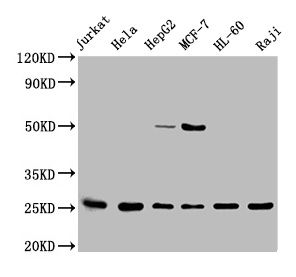
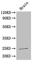
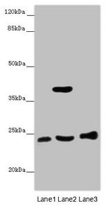
WB analysis of HMGB1 using anti-HMGB1 antibody.Lane 1:human HeLa cell;2:human 293T cell;3:human K562 cell;4:human Jurkat cell;5:rat brain tissue;6:mouse brain tissue.
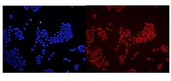
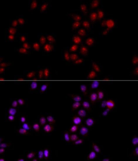
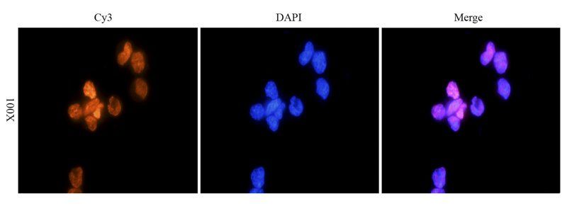
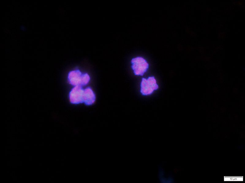
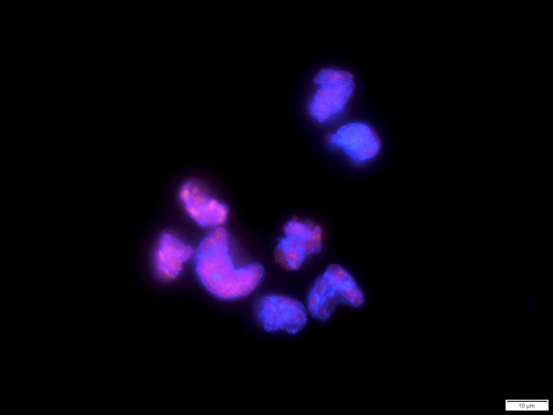
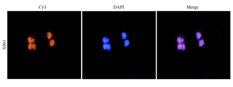
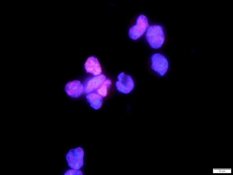
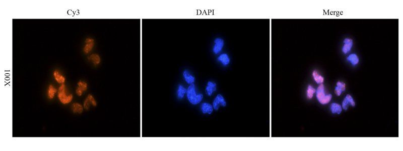
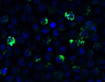
IF analysis of HMGB1 using anti-HMGB1 antibody. HMGB1 was detected in immunocytochemical section of U20S cells.
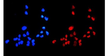
IF analysis of HMGB1 using anti-HMGB1 antibody. HMGB1 was detected in immunocytochemical section of A431 cells.
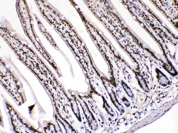
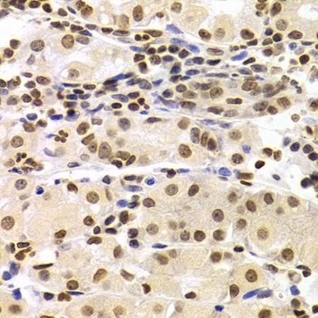
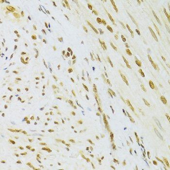
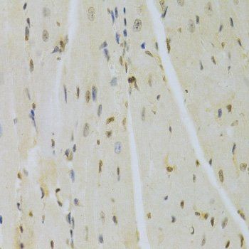
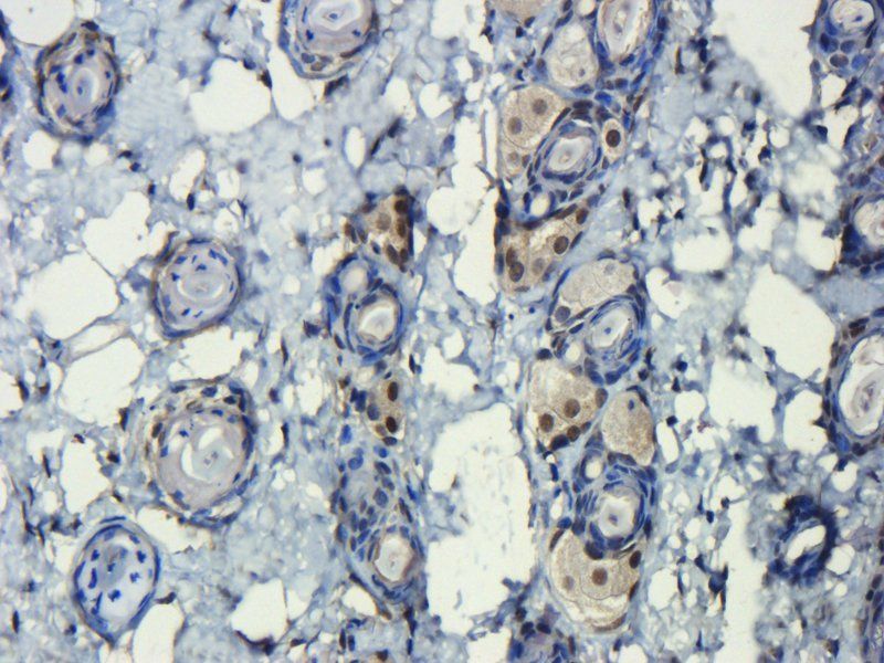
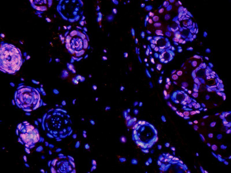
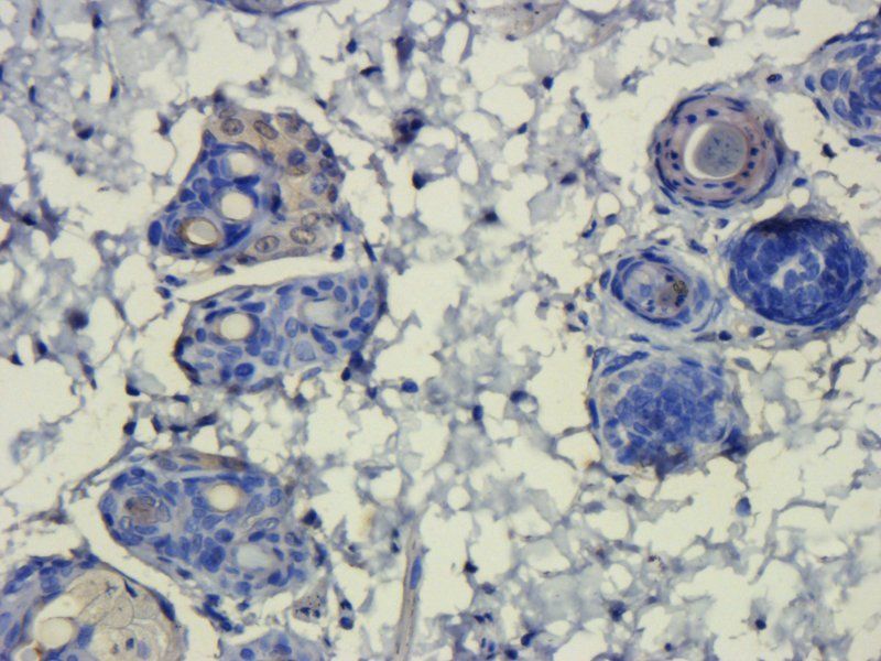
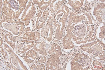
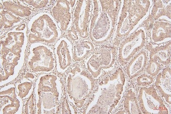
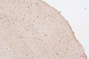
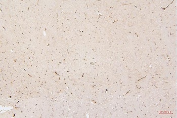
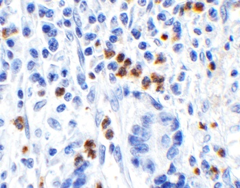
IHC analysis of HMGB1 using anti-HMGB1 antibody. HMGB1 was detected in paraffin-embedded section of mouse intestine tissues.
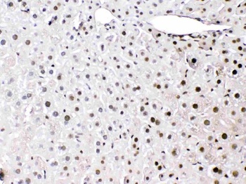
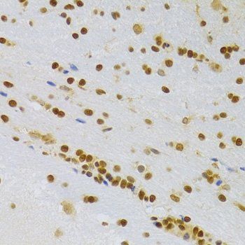
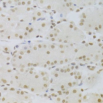
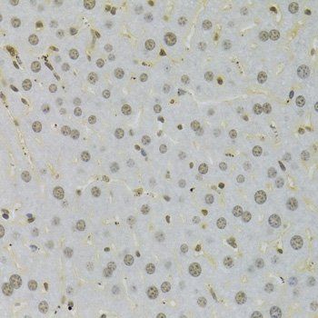
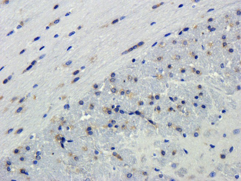
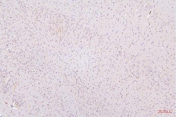
IHC analysis of HMGB1 using anti-HMGB1 antibody. HMGB1 was detected in paraffin-embedded section of mouse liver tissues.
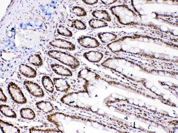
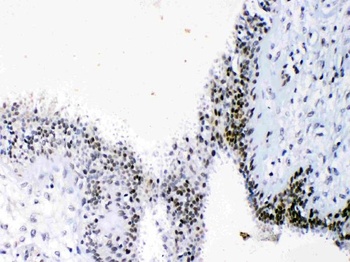
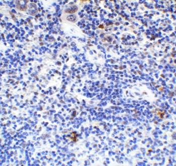
IHC analysis of HMGB1 using anti-HMGB1 antibody. HMGB1 was detected in paraffin-embedded section of rat intestine tissues.
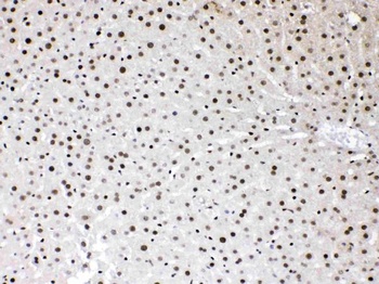
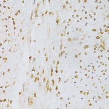
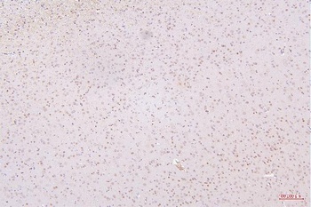
IHC analysis of HMGB1 using anti-HMGB1 antibody. HMGB1 was detected in paraffin-embedded section of rat liver tissues.
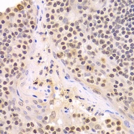
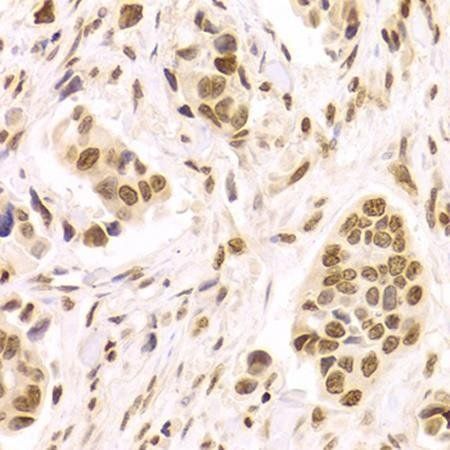
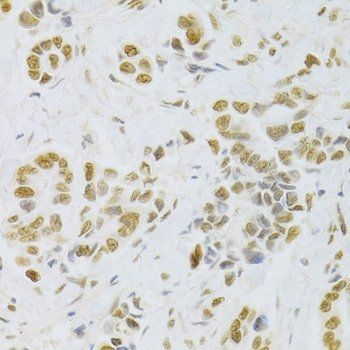
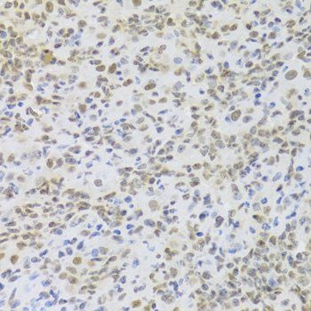
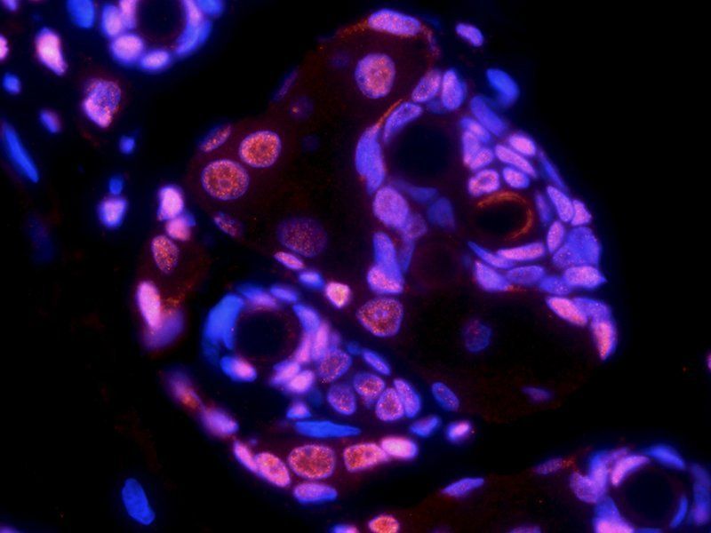
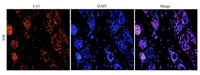
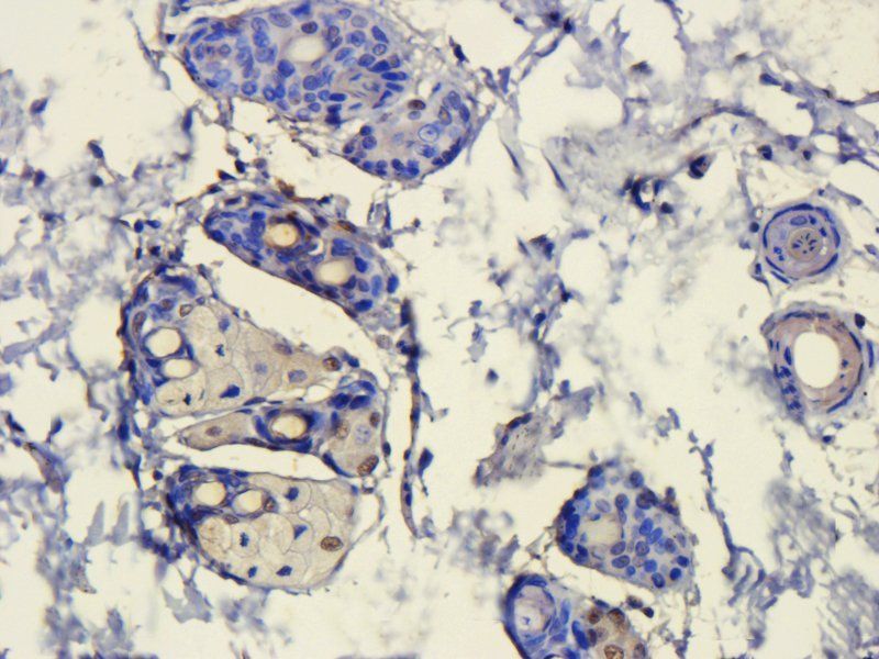
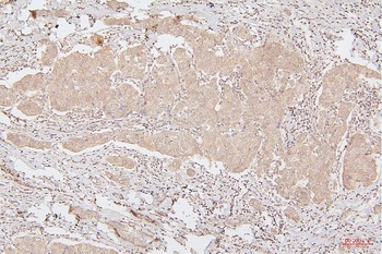
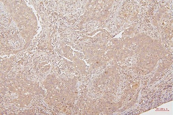
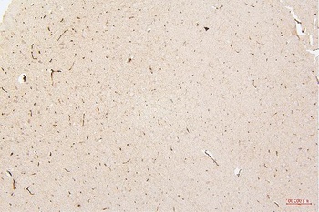
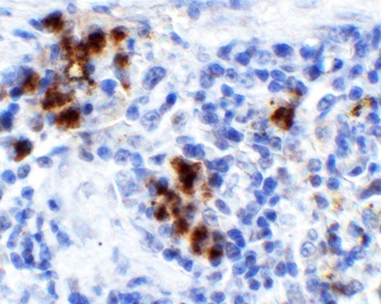
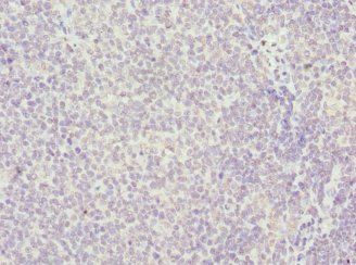
IHC analysis of HMGB1 using anti-HMGB1 antibody. HMGB1 was detected in paraffin-embedded section of human mammary cancer tissues.
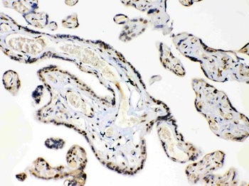
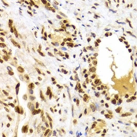
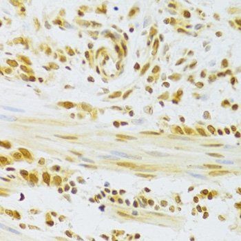
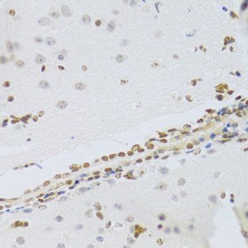
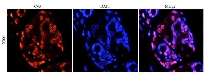
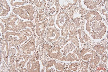
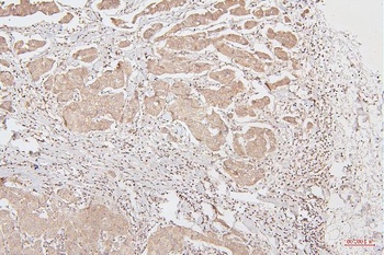
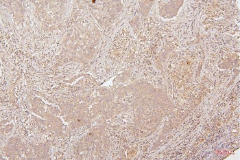
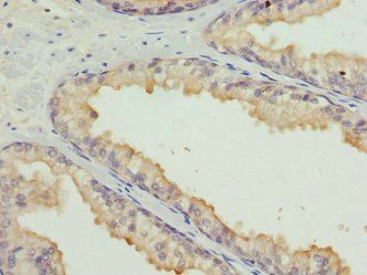
IHC analysis of HMGB1 using anti-HMGB1 antibody. HMGB1 was detected in paraffin-embedded section of human placenta tissues.
- Item 1 of 17
- Item 1 of 16
HMGB1 antibody [orb195321]
ELISA, ICC, IF, IHC-P, WB
Bovine, Human, Mouse, Rat
Rabbit
Polyclonal
Unconjugated
100 μg, 200 μg - Item 1 of 14
HMGB1 Antibody (monoclonal, 5H3) [orb570317]
FC, IHC, WB
Human, Monkey, Mouse, Rat
Mouse
Monoclonal
Unconjugated
10 μg, 100 μg - Item 1 of 8
HMGB1 Antibody [orb1239440]
ELISA, IF, IHC-P, WB
Gallus, Porcine
Human, Mouse, Rat
Rabbit
Polyclonal
Unconjugated
0.1 mg, 0.02 mg - Item 1 of 5

Submit a review
Filter by Rating

- 5 stars

- 4 stars
- 3 stars
- 2 stars
- 1 stars