You have no items in your shopping cart.
Cart summary
Item 1 of 7
Item 1 of 7
HLA-DR/HLA-DRA Antibody (monoclonal, 5B13F7)
Catalog Number: orb1152388

| Catalog Number | orb1152388 |
|---|---|
| Category | Antibodies |
| Description | HLA-DR/HLA-DRA Antibody (monoclonal, 5B13F7) |
| Species/Host | Mouse |
| Clonality | Monoclonal |
| Clone Number | 5B13F7 |
| Tested applications | FC, IHC, WB |
| Reactivity | Human |
| Isotype | Mouse IgG2b |
| Immunogen | E.coli-derived human HLA-DR/HLA-DRA recombinant protein (Position: I26-L254). |
| Concentration | Adding 0.2 ml of distilled water will yield a concentration of 500 μg/ml. |
| Form/Appearance | Lyophilized |
| Conjugation | Unconjugated |
| MW | 35-37 kDa |
| UniProt ID | P01903 |
| Storage | Maintain refrigerated at 2-8°C for up to 2 weeks. For long term storage store at -20°C in small aliquots to prevent freeze-thaw cycles. |
| Note | For research use only |
| Application notes | Tested Species: In-house tested species with positive results. Other applications have not been tested. Optimal dilutions should be determined by end users. Adding 0.2 ml of distilled water will yield a concentration of 500 μg/ml. |
| Expiration Date | 12 months from date of receipt. |
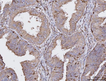
IHC analysis of HLA-DRA using anti-HLA-DRA antibody. HLA-DRA was detected in a paraffin-embedded section of human colonic adenocarcinoma tissue.
IHC analysis of HLA-DRA using anti-HLA-DRA antibody. HLA-DRA was detected in a paraffin-embedded section of human endometrial cancer tissue.
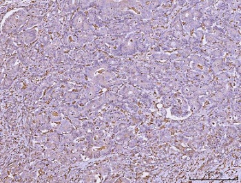
IHC analysis of HLA-DRA using anti-HLA-DRA antibody. HLA-DRA was detected in a paraffin-embedded section of human hepatocellular carcinoma tissue.
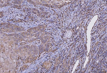
IHC analysis of HLA-DRA using anti-HLA-DRA antibody. HLA-DRA was detected in a paraffin-embedded section of human laryngeal squamous cell carcinoma tissue.
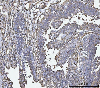
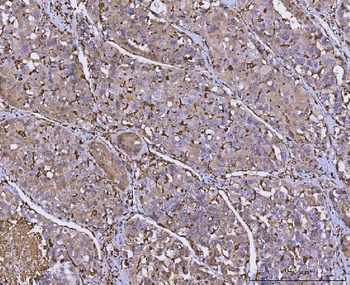
IHC analysis of HLA-DRA using anti-HLA-DRA antibody. HLA-DRA was detected in a paraffin-embedded section of human bladder epithelial carcinoma tissue.
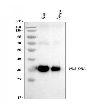
Western blot analysis of HLA-DRA using anti-HLA-DRA antibody.
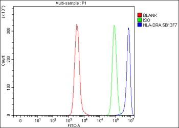
Flow Cytometry analysis of Daudi cells using anti-HLA-DRA antibody(Blue line).Isotype control antibody(Green line) was mouse IgG.Unlabelled sample(Red line) was also used as a control.

Anti-HLA-DR/HLA-DRA Antibody Picoband (monoclonal, 5B13F7) [orb1882117]
FC, IHC, WB
Human
Mouse
Monoclonal
Unconjugated
100 μg
Submit a review
Filter by Rating

- 5 stars

- 4 stars
- 3 stars
- 2 stars
- 1 stars
