You have no items in your shopping cart.
Cart summary
Item 1 of 2
Item 1 of 2
Histone H2A (acetyl K9) HIST1H2AB Monoclonal Antibody
Catalog Number: orb548508

| Catalog Number | orb548508 |
|---|---|
| Category | Antibodies |
| Description | Histone H2A (acetyl K9) HIST1H2AB Monoclonal Antibody |
| Species/Host | Rabbit |
| Clonality | Monoclonal |
| Clone Number | DEB-8 |
| Tested applications | ICC, IF, IHC, WB |
| Reactivity | Human, Mouse, Rat |
| Isotype | Rabbit IgG |
| Immunogen | A synthesized peptide derived from human Histone H2A (acetyl K9) Core component of nucleosome. Nucleosomes wrap and compact DNA into chromatin, limiting DNA accessibility to the cellular machineries which require DNA as a template. Histones thereby play a central role in transcription regulation, DNA repair, DNA replication and chromosomal stability. DNA accessibility is regulated via a complex set of post-translational modifications of histones, also called histone code, and nucleosome remodeling. |
| Concentration | Actual concentration vary by lot. Use suggested dilution ratio to decide dilution procedure. |
| Dilution range | WB 1:500-1:2000IHC 1:500-1:1000ICC/IF 1:500-1:1000 |
| Form/Appearance | Liquid |
| Conjugation | Unconjugated |
| UniProt ID | P04908 |
| Storage | Store at -20°C for one year. For short term storage and frequent use, store at 4°C for up to one month. Avoid repeated freeze-thaw cycles. |
| Note | For research use only |
| Expiration Date | 12 months from date of receipt. |
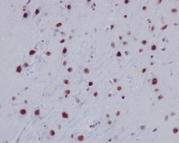
IHC analysis of HIST1H2AB using anti-HIST1H2AB antibody in paraffin-embedded mouse brain (orb548508). HIST1H2AB was detected in paraffin-embedded section. Heat mediated antigen retrieval was performed in citrate buffer (pH6, epitope retrieval solution) for 20 mins. The tissue section was blocked with 10% goat serum. The tissue section was then incubated with 1ug/ml rabbit anti-HIST1H2AB Antibody (orb548508) overnight at 4°C. Biotinylated goat anti-rabbit IgG was used as secondary antibody and incubated for 30 minutes at 37°C. The tissue section was developed using Strepavidin-Biotin-Complex (SABC) (Catalog # orb90444) with DAB as the chromogen.
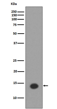
Western blot analysis of HIST1H2AB using anti-HIST1H2AB antibody in HeLa cell lysate treated Trichostatin A. (orb548508). Electrophoresis was performed on a 5-20% SDS-PAGE gel at 70V (Stacking gel)/90V (Resolving gel) for 2-3 hours. The sample well of each lane was loaded with 50ug of sample under reducing conditions. After Electrophoresis, proteins were transferred to a Nitrocellulose membrane at 150mA for 50-90 minutes. Blocked the membrane with 5% Non-fat Milk/TBS for 1.5 hour at RT. The membrane was incubated with rabbit anti-HIST1H2AB antigen affinity purified polyclonal antibody (Catalog # orb548508) at 0.5 ug/mL overnight at 4°C, then washed with TBS-0.1%Tween 3 times with 5 minutes each and probed with a goat anti-rabbit IgG-HRP secondary antibody at a dilution of 1:10000 for 1.5 hour at RT. The signal is developed using an Enhanced Chemiluminescent detection (ECL) kit (Catalog # orb90503) with Tanon 5200 system.

Acetyl-Histone H2A type 1-B/E (K9) antibody [orb418650]
ELISA, ICC, IF
Human
Rabbit
Monoclonal
Unconjugated
100 μl, 50 μlAcetyl-Histone H2A type 1-B/E (K9) Recombinant Antibody [3H5] [orb1995855]
ELISA, ICC, IF
Recombinant
Unconjugated
100 μl
Submit a review
Filter by Rating

- 5 stars

- 4 stars
- 3 stars
- 2 stars
- 1 stars
