You have no items in your shopping cart.
Cart summary
Item 1 of 7
Item 1 of 7
HIST1H2AG Antibody
Catalog Number: orb1265025
| Catalog Number | orb1265025 |
|---|---|
| Category | Antibodies |
| Description | HIST1H2AG Antibody |
| Species/Host | Rabbit |
| Clonality | Polyclonal |
| Tested applications | FC, IHC-P, WB |
| Predicted Reactivity | Bovine, Drosophila, Gallus, Monkey, Rat, Xenopus, Yeast, Zebrafish |
| Reactivity | Human, Mouse |
| Isotype | Rabbit Ig |
| Immunogen | This HIST1H2AG antibody is generated from a rabbit immunized with a KLH conjugated synthetic peptide between 63-87 amino acids from the Central region of human HIST1H2AG. |
| Concentration | batch dependent |
| Dilution range | For WB starting dilution is: 1:2000For IHC-P starting dilution is: 1:25For FACS starting dilution is: 1:25 |
| Form/Appearance | Liquid |
| Conjugation | Unconjugated |
| MW | 14 kDa |
| Target | HIST1H2AG |
| UniProt ID | P0C0S8 |
| NCBI | P0C0S8 |
| Storage | Store at 4°C for three months and -20°C, stable for up to one year. As with all antibodies care should be taken to avoid repeated freeze thaw cycles. Antibodies should not be exposed to prolonged high temperatures. |
| Buffer/Preservatives | Supplied in PBS with 0.09% (W/V) sodium azide. |
| Alternative names | Histone H2A type 1, H2A1, Histone H2A/p, HIST1H2AG Read more... |
| Note | For research use only |
| Application notes | For WB starting dilution is: 1:2000For IHC-P starting dilution is: 1:25For FACS starting dilution is: 1:25 |
| Expiration Date | 12 months from date of receipt. |
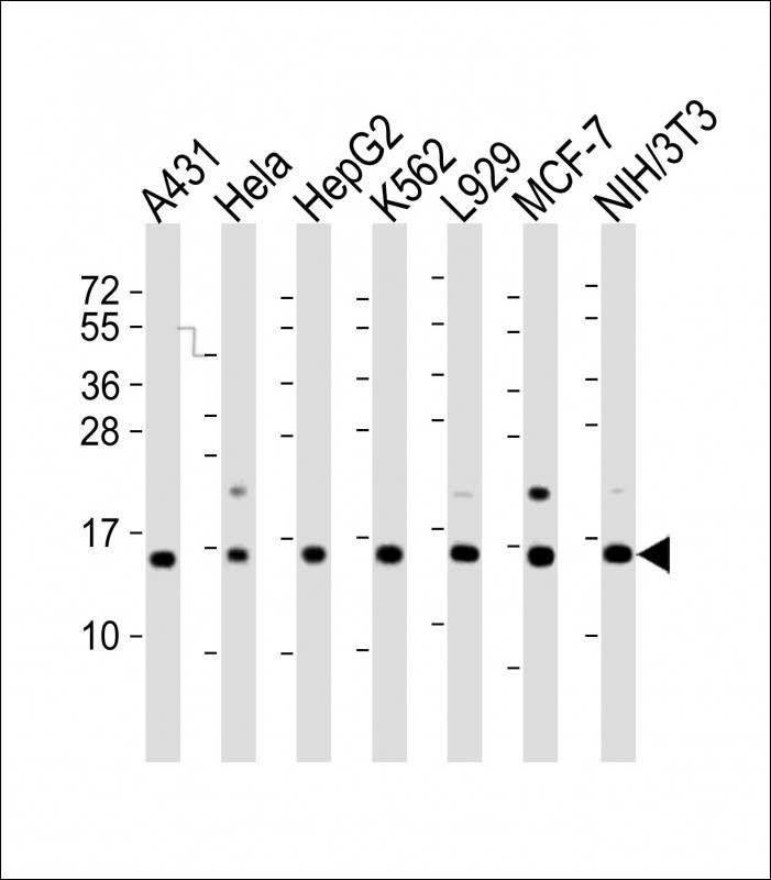
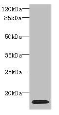
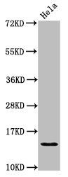
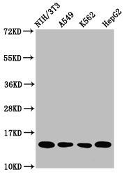
Western Blot at 1:2000 dilution Lane 1: A431 whole cell lysate Lane 2: Hela whole cell lysate Lane 3: HepG2 whole cell lysate Lane 4: K562 whole cell lysate Lane 5: L929 whole cell lysate Lane 6: MCF-7 whole cell lysate Lane 7: NIH/3T3 whole cell lysate Lysates/proteins at 20 ug per lane.
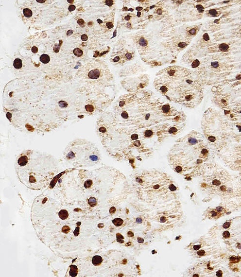
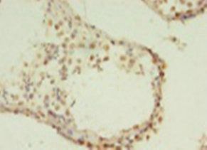
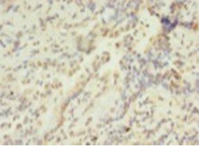
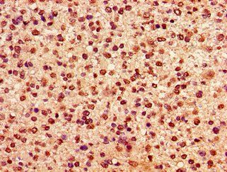
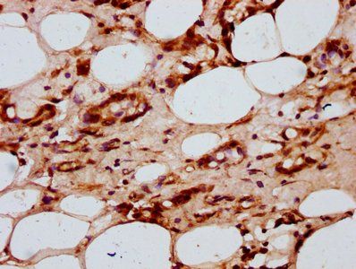
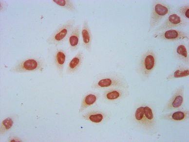
Immunohistochemical analysis of paraffin-embedded M. pancreas section using HIST1H2AG Antibody. Antibody was diluted at 1:100 dilution. A peroxidase-conjugated goat anti-rabbit IgG at 1:400 dilution was used as the secondary antibody, followed by DAB staining.
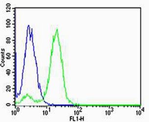
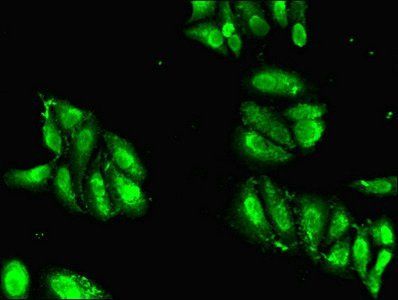
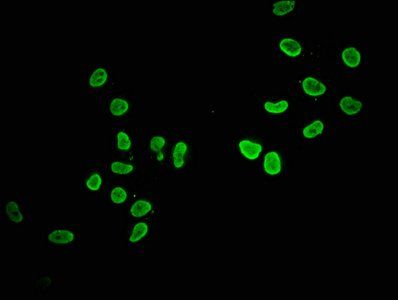
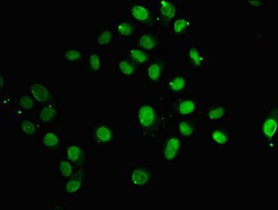
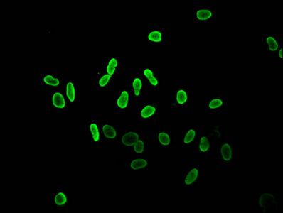
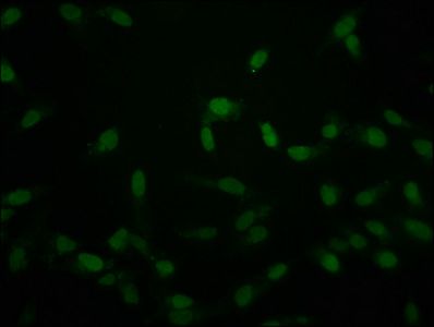
Flow cytometric analysis of Hela cells using HIST1H2AG Antibody (green) compared to an isotype control of rabbit IgG (blue). Antibody was diluted at 1:25 dilution. An Alexa Fluor 488 goat anti-rabbit lgG at 1:400 dilution was used as the secondary antibody.
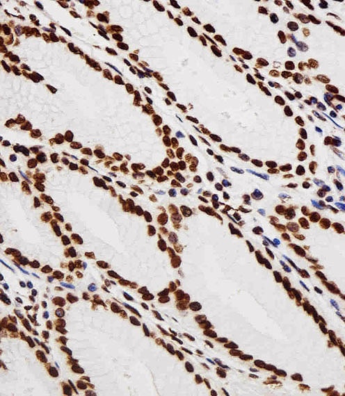
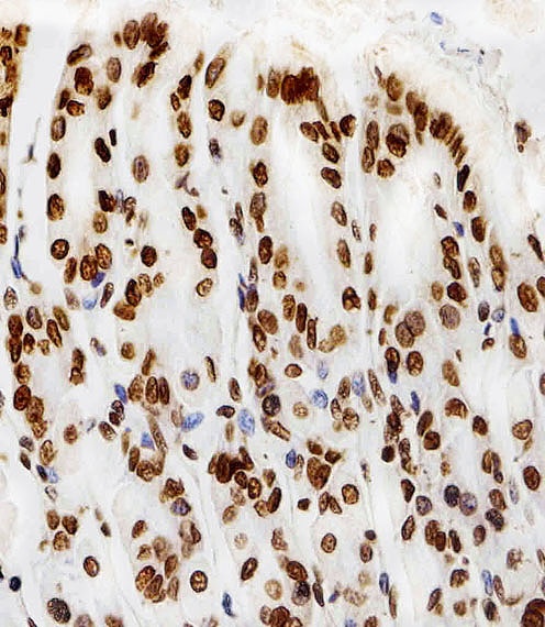
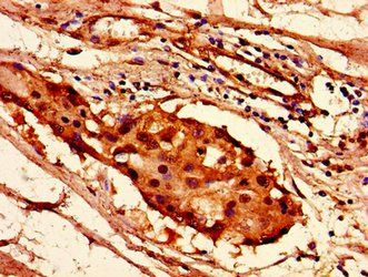
Immunohistochemical analysis of paraffin-embedded M. testis section using H. stomach Antibody. Antibody was diluted at 1:100 dilution. A peroxidase-conjugated goat anti-rabbit IgG at 1:400 dilution was used as the secondary antibody, followed by DAB staining.
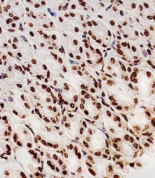
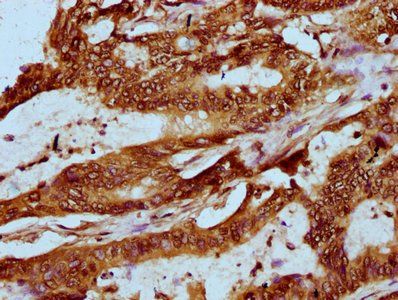
Immunohistochemical analysis of paraffin-embedded R. stomach section using HIST1H2AG Antibody. Antibody was diluted at 1:100 dilution. A peroxidase-conjugated goat anti-rabbit IgG at 1:400 dilution was used as the secondary antibody, followed by DAB staining.
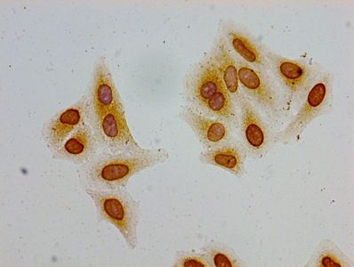
Immunohistochemical analysis of paraffin-embedded M. stomach section using HIST1H2AG Antibody. Antibody was diluted at 1:100 dilution. A peroxidase-conjugated goat anti-rabbit IgG at 1:400 dilution was used as the secondary antibody, followed by DAB staining.
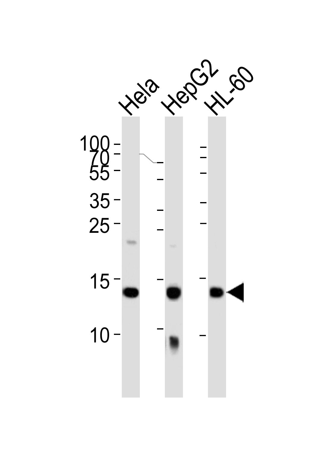
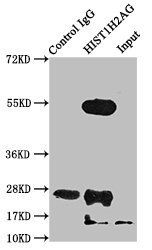
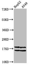
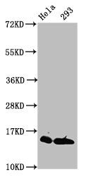
Western blot analysis of lysates from Hela, HepG2, HL-60 cell line (from left to right), using HIST1H2AG Antibody. was diluted at 1:1000 at each lane.
- Item 1 of 5
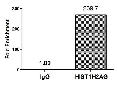
HIST1H2AG antibody [orb350509]
ChIP, ELISA, IF, IHC, WB
Human
Rabbit
Polyclonal
Unconjugated
100 μg, 50 μg - Item 1 of 5
HIST1H2AG (Ab-36) antibody [orb416611]
ChIP, ELISA, IF, IHC, WB
Human
Rabbit
Polyclonal
Unconjugated
100 μl, 50 μl - Item 1 of 5
HIST1H2AG (Ab-118) antibody [orb517097]
ELISA, IF, IHC, IP, WB
Human, Mouse
Rabbit
Polyclonal
Unconjugated
50 μl, 100 μl - Item 1 of 4
Acetyl-HIST1H2AG (K36) antibody [orb416601]
ChIP, ELISA, ICC, IF, WB
Human
Rabbit
Polyclonal
Unconjugated
50 μl, 100 μl - Item 1 of 4
Crotonyl-HIST1H2AG (K118) antibody [orb416650]
ChIP, ELISA, ICC, IF, WB
Human
Rabbit
Polyclonal
Unconjugated
100 μl, 50 μl
Submit a review
Filter by Rating

- 5 stars

- 4 stars
- 3 stars
- 2 stars
- 1 stars