You have no items in your shopping cart.
Cart summary
Item 1 of 2
Item 1 of 2
HDAC8/Histone Deacetylase 8 Rabbit Monoclonal Antibody
Catalog Number: orb547876
| Catalog Number | orb547876 |
|---|---|
| Category | Antibodies |
| Description | HDAC8/Histone Deacetylase 8 Rabbit Monoclonal Antibody |
| Species/Host | Rabbit |
| Clonality | Monoclonal |
| Clone Number | HGO-8 |
| Tested applications | FC, ICC, IF, IP, WB |
| Reactivity | Human, Mouse, Rat |
| Isotype | Rabbit IgG |
| Immunogen | A synthesized peptide derived from human HDAC8 |
| Concentration | Actual concentration vary by lot. Use suggested dilution ratio to decide dilution procedure. |
| Dilution range | WB 1:5000-1:20000 IP 1:50 Flow Cytometry 1:50 |
| Form/Appearance | Liquid |
| Conjugation | Unconjugated |
| MW | 41758 MW |
| UniProt ID | Q9BY41 |
| Storage | Store at -20°C for one year. For short term storage and frequent use, store at 4°C for up to one month. Avoid repeated freeze-thaw cycles. |
| Alternative names | Histone deacetylase 8;HD8;3.5.1.98;HDAC8;HDACL1;CD Read more... |
| Note | For research use only |
| Expiration Date | 12 months from date of receipt. |
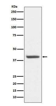
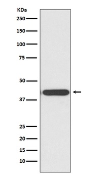
Western blot analysis of HDAC8 expression in HeLa cell lysate (orb547876). Electrophoresis was performed on a 5-20% SDS-PAGE gel at 70V (Stacking gel)/90V (Resolving gel) for 2-3 hours. The sample well of each lane was loaded with 50ug of sample under reducing conditions. After Electrophoresis, proteins were transferred to a Nitrocellulose membrane at 150mA for 50-90 minutes. Blocked the membrane with 5% Non-fat Milk/TBS for 1.5 hour at RT. The membrane was incubated with rabbit anti-HDAC8 monoclonal antibody (Catalog # orb547876) overnight at 4°C, then washed with TBS-0.1%Tween 3 times with 5 minutes each and probed with a goat anti-rabbit IgG-HRP secondary antibody at a dilution of 1:10000 for 1.5 hour at RT. The signal is developed using an Enhanced Chemiluminescent detection (ECL) kit (Catalog # orb90503) with Tanon 5200 system. A specific band was detected for HDAC8.
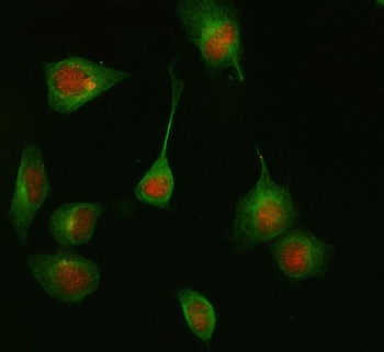
IF analysis of HDAC8 using anti-HDAC8 antibody (orb547876) and anti-Beta Tubulin antibody (orb623835). HDAC8 was detected in immunocytochemical section of HELA cell. Enzyme antigen retrieval was performed using IHC enzyme antigen retrieval reagent (orb90553) for 15 mins. The cells were blocked with 10% goat serum. And then incubated at 1: 50 with rabbit anti-HDAC8 Antibody (orb547876) and mouse anti-Beta Tubulin antibody (orb623835) overnight at 4°C. Cy3 Conjugated Goat Anti-Rabbit IgG (orb27705) and DyLight®488 Conjugated Goat Anti-Mouse IgG were used as secondary antibody at 1: 500 dilution and incubated for 30 minutes at 37°C. Visualize using a fluorescence microscope and filter sets appropriate for the label used.
- Item 1 of 1
HDAC8/Histone Deacetylase 8 Rabbit Monoclonal Antibody [orb547875]
FC, ICC, IF, IHC, IP, WB
Human
Rabbit
Monoclonal
Unconjugated
30 μl, 100 μl
Submit a review
Filter by Rating

- 5 stars

- 4 stars
- 3 stars
- 2 stars
- 1 stars