You have no items in your shopping cart.
Cart summary
Item 1 of 6
Item 1 of 6
HDAC2 Antibody (C-term)
Catalog Number: orb1938513

| Catalog Number | orb1938513 |
|---|---|
| Category | Antibodies |
| Description | Purified Rabbit Polyclonal Antibody (Pab) |
| Target | This HDAC2 antibody is generated from rabbits immunized with a KLH conjugated synthetic peptide between 456-488 amino acids from the C-terminal region of human HDAC2. |
| Clonality | Polyclonal |
| Species/Host | Rabbit |
| Isotype | Rabbit IgG |
| Conjugation | Unconjugated |
| Reactivity | Human |
| Form/Appearance | Purified polyclonal antibody supplied in PBS with 0.09% (W/V) sodium azide. This antibody is prepared by Saturated Ammonium Sulfate (SAS) precipitation followed by dialysis against PBS. |
| UniProt ID | Q92769 |
| MW | 55364 Da |
| Tested applications | IF, IHC-P, WB |
| Dilution range | IF: 1:10~50, IF: 1:10~50, WB: 1:1000, WB: 1:1000, WB: 1:1000, IHC-P: 1:10~50 |
| Antibody Type | Primary Antibody |
| Clone Number | RB5718 |
| Storage | Maintain refrigerated at 2-8°C for up to 2 weeks. For long term storage store at -20°C in small aliquots to prevent freeze-thaw cycles |
| Alternative names | Histone deacetylase 2, HD2, HDAC2 |
| Note | For research use only |
| NCBI | NP_001518.3 |
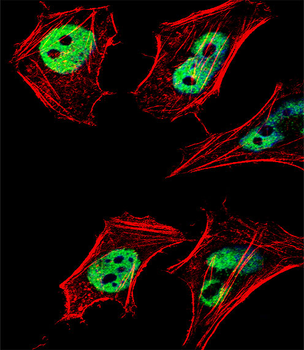
Confocal immunofluorescent analysis of HDAC2 Antibody (C-term) with 293 cell followed by Alexa Fluor 488-conjugated goat anti-rabbit lgG (green). Actin filaments have been labeled with Alexa Fluor 555 phalloidin (red).
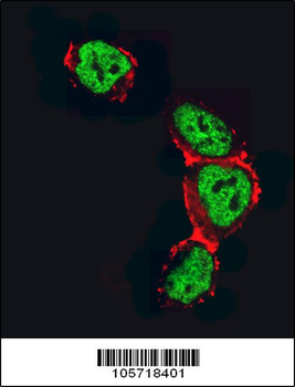
Fluorescent confocal image of Hela cell stained with HDAC2 Antibody (C-term).Hela cells were fixed with 4% PFA (20 min), permeabilized with Triton X-100 (0.1%, 10 min), then incubated with HDAC2 primary antibody (1:25, 1 h at 37°C). For secondary antibody, Alexa Fluor 488 conjugated donkey anti-rabbit antibody (green) was used (1:400, 50 min at 37°C).Cytoplasmic actin was counterstained with Alexa Fluor 555 (red) conjugated Phalloidin (7units/ml, 1 h at 37°C). Nuclei were counterstained with DAPI (blue) (10 µg/ml, 10 min). hHDAC2 immunoreactivity is localized to Nucleus significantly.
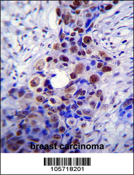
HDAC2 Antibody (C-term) immunohistochemistry analysis in formalin fixed and paraffin embedded human breast carcinoma followed by peroxidase conjugation of the secondary antibody and DAB staining. This data demonstrates the use of HDAC2 Antibody (C-term) for immunohistochemistry. Clinical relevance has not been evaluated.
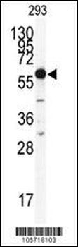
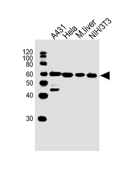
Western blot analysis of anti-HDAC2 Antibody (C-term) in 293 cell line lysates (35 ug/lane). HDAC2 (arrow) was detected using the purified Pab.

Western blot analysis of anti-HDAC2 Antibody (C-term) in mouse spleen tissue lysates (35 ug/lane). HDAC2 (arrow) was detected using the purified Pab.
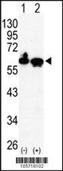
Western blot analysis of HDAC2 (arrow) using rabbit polyclonal HDAC2 Antibody (C-term).293 cell lysates (2 ug/lane) either nontransfected (Lane 1) or transiently transfected with the HDAC2 gene (Lane 2).
- Item 1 of 4