You have no items in your shopping cart.
Cart summary
Item 1 of 5
Item 1 of 5
HDAC-1 antibody
Catalog Number: orb345508
| Catalog Number | orb345508 |
|---|---|
| Category | Antibodies |
| Description | HDAC-1 antibody |
| Species/Host | Rabbit |
| Clonality | Polyclonal |
| Tested applications | ELISA, IF, IHC, Multiplex Assay, WB |
| Reactivity | Human |
| Isotype | IgG |
| Immunogen | Anti-HDAC-1 antibody was prepared from whole rabbit serum produced by repeated immunizations with a synthetic peptide corresponding to a C-Terminal region near amino acids 450-482 of Human HDAC-1. |
| Concentration | 1.33 mg/mL |
| Dilution range | ELISA: 1:10,000 - 1:50,000, IHC: 1:200 - 1:1,000, IF: 10ug / ml, WB: 1:1,000 - 1:5,000 |
| Form/Appearance | Liquid (sterile filtered) |
| Purity | Anti-HDAC-1 antibody is directed against human HDAC-1 protein. HDAC-1 antibody was affinity purified from monospecific antiserum by immunoaffinity purification. A BLAST analysis was used to suggest reactivity with this protein from human, mouse, rat and chimpanzee sources based on 100% homology for the immunogen sequence. Cross reactivity may occur with HDAC-1 from bovine (82% homology) and chicken (80% homology) sources. Cross reactivity with HDAC-1 homologues from other sources has not been determined. |
| Conjugation | Unconjugated |
| UniProt ID | Q13547 |
| NCBI | 13128860 |
| Storage | Store vial at -20° C prior to opening. Aliquot contents and freeze at -20° C or below for extended storage. Avoid cycles of freezing and thawing. Centrifuge product if not completely clear after standing at room temperature. This product is stable for several weeks at 4° C as an undiluted liquid. Dilute only prior to immediate use. |
| Buffer/Preservatives | 0.01% (w/v) Sodium Azide |
| Alternative names | rabbit anti-HDAC-1 antibody, HDAC1, HDAC 1, HD 1 a Read more... |
| Note | For research use only |
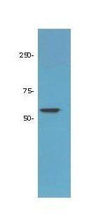
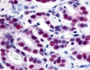
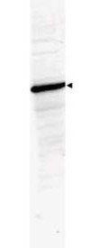
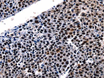
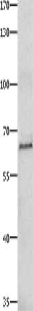
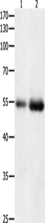
| Application notes | Anti-HDAC-1 Antibody has been tested for use in ELISA, immunohistochemistry, immunofluorescence, and western blot. Specific conditions for reactivity should be optimized by the end user. Specific nuclear staining is observed by IHC. Expect bands at 65 kDa in size corresponding to HDAC-1 by western blotting in the appropriate cell lysate or extract. |
| Expiration Date | 12 months from date of receipt. |
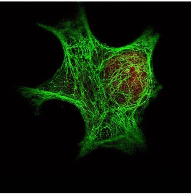
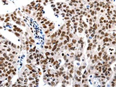
Immunofluorescence analysis of used with Dylight 488 (shown in green) to detect Keratin. Data was collected on a STED-CW TCS-SP5 Confocal system (Leica Microsystems). using HDAC-1 antibody
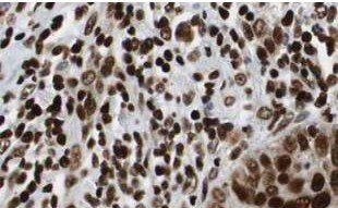
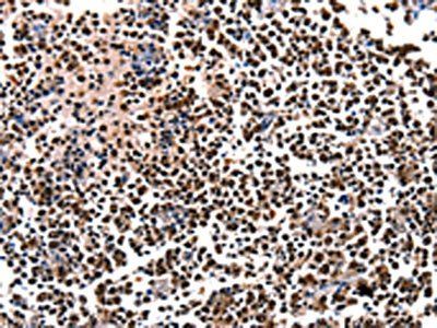
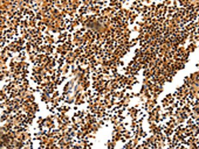
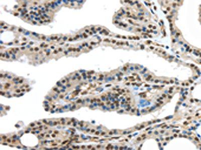
Immunohistochemical staining of human lung tissue using HDAC-1 antibody
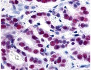
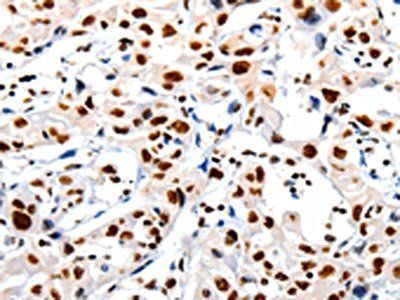
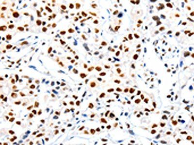
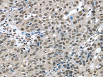
Immunohistochemical staining of human prostate cancer tissue using HDAC-1 antibody

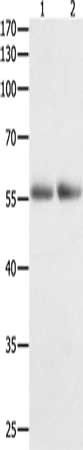
Western blot analysis of Lane 1: 293 whole cell lysate. Lane 2: none. Load: 35 ug per lane using HDAC-1 antibody
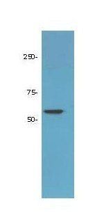
Western blot analysis of Lane 1: LNCaP prostate cancer cells. Lane 2: none. Load: 50 ug per lane using HDAC-1 antibody
- Item 1 of 5
HDAC-1 antibody [orb345509]
ELISA, IF, IHC, Multiplex Assay, WB
Human
Rabbit
Polyclonal
Unconjugated
25 μl - Item 1 of 3
HDAC1 antibody [orb519005]
ELISA, IHC, WB
Human, Mouse, Rat
Rabbit
Polyclonal
Unconjugated
50 μl, 100 μl - Item 1 of 3
HDAC1 antibody [orb522563]
ELISA, IHC, WB
Human, Mouse, Rat
Rabbit
Polyclonal
Unconjugated
100 μl, 50 μl - Item 1 of 3
HDAC1 antibody [orb522564]
ELISA, IHC, WB
Human, Mouse, Rat
Rabbit
Polyclonal
Unconjugated
100 μl, 50 μl - Item 1 of 2


Submit a review
Filter by Rating

- 5 stars

- 4 stars
- 3 stars
- 2 stars
- 1 stars