You have no items in your shopping cart.
Cart summary
Item 1 of 1
HCG-beta Antibody Cocktail
Catalog Number: orb385743
| Catalog Number | orb385743 |
|---|---|
| Category | Antibodies |
| Description | This mAb reacts with a protein of 22kDa, identified as the beta sub-unit of HCG. It does not cross react with the alpha sub-unit. HCG is a glycoprotein, which is secreted in large quantities by normal trophoblasts. It is present only in trace amounts in non-pregnant urine and sera but rises sharply during pregnancy. HCG is composed of two non-identical, non-covalently linked polypeptide chains designated as the alpha and beta subunits. The alpha subunit is identical to that of thyroid stimulating hormone (TSH), follicle stimulating hormone (FSH), and luteinizing hormone (LH). hCG mAb detects cells and tumors of trophoblastic origin such as choriocarcinoma. Large cell carcinoma and adenocarcinoma of the lung demonstrate anti-hCG positivity in 90% and 60% of cases respectively. 20% of lung squamous cell carcinomas are positive. hCG expression by non-trophoblastic tumors may indicate aggressive behavior. |
| Species/Host | Mouse |
| Clonality | Monoclonal |
| Clone Number | HCGb/54 + HCGb/459 |
| Tested applications | IHC-P |
| Reactivity | Human |
| Isotype | Mouse IgG1, kappa |
| Immunogen | Recombinant hCG beta protein was used as the immunogens for this hCG beta antibody cocktail. |
| Dilution range | Immunohistochemistry (FFPE): 0.5-1ug/ml for 30 min at RT |
| Purity | Protein G affinity chromatography |
| Conjugation | Unconjugated |
| Formula | 0.2 mg/ml in 1X PBS with 0.1 mg/ml BSA (US sourced) and 0.05% sodium azide |
| Hazard Information | This HCG-beta antibody cocktail is available for research use only. |
| UniProt ID | P01233 |
| Storage | Store the HCG-beta antibody cocktail at 2-8°C (with azide) or aliquot and store at -20°C or colder (without azide). |
| Buffer/Preservatives | 0.2 mg/ml in 1X PBS with 0.1 mg/ml rAlbumin (US sourced) and 0.05% sodium azide |
| Note | For research use only |
| Application notes | The optimal dilution of the HCG-beta antibody for each application should be determined by the researcher.1. Staining of formalin-fixed tissues requires boiling tissue sections in 10mM citrate buffer, pH 6.0, for 10-20 min followed by cooling at RT for 20 minutes.2. The prediluted format is supplied in a dropper bottle and is optimized for use in IHC. After epitope retrieval step (if required), drip mAb solution onto the tissue section and incubate at RT for 30 min. |
| Expiration Date | 12 months from date of receipt. |
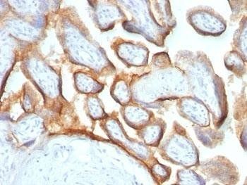
Immunohistochemical staining of Human placenta using HCG-beta Cocktail antibody
Submit a review
Filter by Rating

- 5 stars

- 4 stars
- 3 stars
- 2 stars
- 1 stars