You have no items in your shopping cart.
Description
Research Area
Protein Biochemistry
Images & Validation
−Item 1 of 5
| Tested Applications | SDS-PAGE |
|---|---|
| Application Notes |
Key Properties
−| Source | Golden Syrian Hamster |
|---|---|
| Biological Origin | Golden Syrian Hamster |
| Isotype | IgG |
| Conjugation | Unconjugated |
| Purity | Hamster IgG whole molecule was prepared from normal serum by a multi-step process which includes delipidation, salt fractionation and ion exchange chromatography followed by extensive dialysis against the buffer stated above. Hamster IgG whole molecule was assayed by immunoelectrophoresis resulted in a single precipitin arc against anti-GS Hamster IgG and anti-GS Hamster Serum. |
Storage & Handling
−| Storage | Store vial at 4° C prior to restoration. For extended storage aliquot contents and freeze at -20° C or below. Avoid cycles of freezing and thawing. Centrifuge product if not completely clear after standing at room temperature. Hamster IgG whole molecule is stable for several weeks at 4° C as an undiluted liquid. Dilute only prior to immediate use. |
|---|---|
| Form/Appearance | Lyophilized |
| Buffer/Preservatives | Preservative: 0.01% (w/v) Sodium Azide; Buffer: 0.02 M Potassium Phosphate, 0.15 M Sodium Chloride, pH 7.2 |
| Concentration | 10.0 mg/mL |
| Expiration Date | 12 months from date of receipt. |
| Disclaimer | For research use only |
Alternative Names
−Hamster Immunoglobulin Gamma, Immunoglobulin G
Similar Products
−
Rabbit Hamster IgG (heavy and light chains), conjugated with FITC Antibody [orb21619]
Hamster
Rabbit
Polyclonal
FITC
2 mlGoat IgG Hamster IgG (heavy and light chains), conjugated with FITC Antibody [orb22040]
Hamster
Goat
Polyclonal
FITC
2 mlGoat Hamster IgG (heavy and light chains) Antibody [orb22055]
Hamster
Goat
Polyclonal
Unconjugated
1 mlRabbit Hamster IgG (heavy and light chains), conjugated with Horseradish peroxidase Antibody [orb21618]
Hamster
Rabbit
Polyclonal
HRP
1 mlGoat IgG Hamster IgG (heavy and light chains), conjugated with Horseradish peroxidase Antibody [orb22039]
DOT, ELISA, ICC, WB
Hamster
Goat
Polyclonal
HRP
1 ml
Quality Guarantee
Explore bioreagents carefree to elevate your research. All our products are rigorously tested for performance. If a product does not perform as described on its datasheet, our scientific support team will provide expert troubleshooting, a prompt replacement, or a refund. For full details, please see our Terms & Conditions and Buying Guide. Contact us at [email protected].
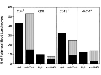
Anti-CD154–facilitated alloengraftment is multilineage. Twenty mice from 2 representative experiments shown in Figure 1 were phenotyped at 120 days after BMT for donor-host origin of CD4+ and CD8 + T cells, CD19 + B cells, and MAC-1 + myeloid cells. On the x-axis are shown the host and donor proportions of each of the lineages. ▪ indicates the proportion of each lineage of host origin; ▥, the proportion of each lineage that is of donor origin. On the y-axis is shown the percentage of PBLs of each lineage. Irrelevant hIgG–treated mice had no detectable donor chimerism and thus are composed entirely of host-type cells. Note that most CD4+ T cells, CD19 + B cells, and MAC-1 + myeloid cells in anti-CD154–treated mice are of donor origin. In contrast, most of the CD8 + T cells are of host origin.
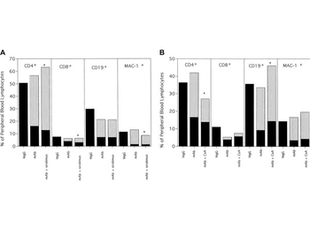
Anti-CD40L mAb alone or in combination with (A) sirolimus or (B) CsA results in long-term multilineage engraftment.
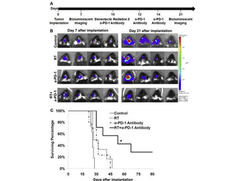
Anti-PD-1 antibody plus radiation therapy (RT) cures mice with intracranial GL261-luc tumors. (A) Experimental timeline. (B) Luciferase imaging of 4 distinct mice per treatment arm before treatment (day 7) and after treatment (day 21), divided by treatment group. All images at same scale. All mice individually matched on days 7 and 21. (C) Kaplan-Meier survival curve. P 6 mice per arm.
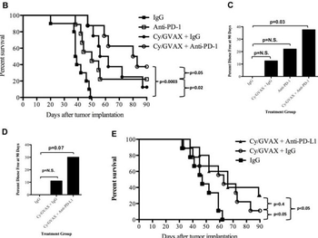
Combination therapy with Cy/GVAX and PD-1 or PD-L1 blockade improves clinical outcomes in a PDA mouse model. Anti-PD-1, anti-PD-L1 or IgG (5 mg/kg IP) were administered IP twice weekly until death starting on day 3. (B) Kaplan-Meier survival curves of mice that were implanted with PDA cells and were treated with different combinations of Cy, GVAX and the αPD-1 antibody. The percentages of mice that remained disease free at day 90 following tumor implantation and therapy with (C) Cy, GVAX and/or αPD-1 or (D) Cy, GVAX and αPD-L1 are shown. All the p values were yielded by comparing GVAX and/or αPD-1/αPD-L1 treatment groups with IgG treated group. (E) Kaplan-Meier survival curves of mice that were implanted with Panc02 cells via hemispleen technique and treated with different combinations of Cy, GVAX and αPD-L1 antibody. Data are represented as results obtained from experiments with 8-10 mice per group that were repeated at least twice.
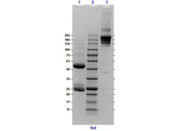
SDS-PAGE Results of Golden Syrian Hamster IgG Whole Molecule. Lane 1: Golden Syrian Hamster IgG Whole Molecule, Reduced [5.0 µg]. Lane 2: Opal Pre-Stained Molecular Weight Marker. Lane 3: Golden Syrian Hamster IgG Whole Molecule, Non-Reduced [5.0 µg]. 4-20% gel, Coomassie stained.
Documents Download
Datasheet
Product Information
Request a Document
Protocol Information
Protein Handling and Storage Guide
Protein Handling Guide
SDS-PAGE
Sodium Dodecyl Sulphate PolyAcrylamide Gel Electrophoresis
Hamster IgG Antibody (orb346158)

- 0.0
Based on 0 reviews
Participating in our Biorbyt product reviews program enables you to support fellow scientists by sharing your firsthand experience with our products.
Login to Submit a ReviewAvailable Sizes
Select a size below