You have no items in your shopping cart.
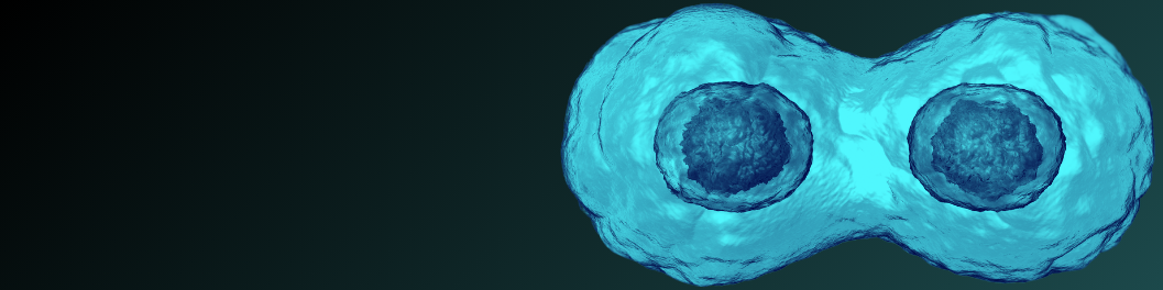
What is cell proliferation?
Cell proliferation is the increase in the number of cells as a result of cell division. The study of cell proliferation is important for the understanding of healthy cellular processes and is also a benchmark for studying the nature and progression of a number of diseases.
Phases of the cell cycle
The cell cycle is composed of 2 major phases - interphase and mitosis. During interphase, the cell grows and replicates DNA. During mitosis, the cell equally distributes chromosomes and divides into two cells. This process is visualized in the diagrams below.

Clinical Relevance
The study of cell proliferation is clinically relevant in a number of areas, including:
- Developmental biology
- Diagnostics
- Wound healing and tissue repair
- Cancer research
- Regenerative medicine
- Development of therapeutic pharmaceuticals
How scientists measure cell proliferation
Nucleoside-Analog Incorporation Assays
In this method, scientists introduce radioactive or chemically tagged nucleosides into the cell during the S phase of interphase, in which DNA replication occurs. Scientists can then identify and analyze cells with these labeled nucleosides
- Tritiated thymidine ([3H]TdR) incorporation assay: In this assay, radiolabeled thymidine is added to cell cultures, given time to proliferate, and measured by a liquid scintillation counter.
- BrdU cell proliferation assay: In this assay, 5-bromo-2′-deoxyuridine (BrdU), a thymidine analog, is introduced into cells. These cells can then be identified through the binding of a fluorescently tagged BrdU specific antibody or through a secondary antibody.
Cell Proliferation Proteins
Scientists can also use proteins to identify cell proliferation. There are particular proteins that occur exclusively during cell proliferation that are absent from non-proliferating cells. Scientists can use antibodies to identify the presence of these proteins. Protein detection offers many advantages, the largest being the ability to use multiple techniques (ie. microscopy, IF, WB) for detection.
- Ki-67: This is the most commonly assayed protein for cell proliferation. Ki-67 is expressed during interphase and mitosis.
- Histone H3:A cell proliferating protein expressed during mitosis.
- Proliferating cell nuclear antigen (PCNA):A cell proliferating protein that peaks during the S phase of interphase.
Cytoplasmic Proliferation Dyes
In this method of analysis, scientists use a fluorescent dye that binds to the cytoplasmic components of cells. Scientists then measure the fluorescence intensity, as two daughter cells will each have a fluorescence that is half the intensity of its former parent cell. Carboxyfluorescein diacetate succinimidyl ester (CFSE), a green fluorescent dye, is the main dye used for this technique.
Counting
- Manual counting: In this method, scientists stain cells with a dye to differentiate living from dead cells. Trypan blue is one of the most common dyes used for this purpose and allows for differentiation by staining non-living cells that have damaged cell membranes.
- Automated counting: In this method, scientists use automated cell counters, which provides a more accurate result than manual counting.
References
- George A. Romar, Thomas S. Kupper, Sherrie J. Divito, Research Techniques Made Simple: Techniques to Assess Cell Proliferation, Journal of Investigative Dermatology, Volume 136, Issue 1, 2016, Pages e1-e7, ISSN 0022-202X, https://doi.org/10.1016/j.jid.2015.11.020.