You have no items in your shopping cart.
Cart summary
Item 1 of 4
Item 1 of 4
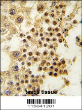
GDF3 Antibody
Catalog Number: orb1272739
| Catalog Number | orb1272739 |
|---|---|
| Category | Antibodies |
| Description | GDF3 Antibody |
| Species/Host | Rabbit |
| Clonality | Polyclonal |
| Tested applications | ELISA, WB |
| Reactivity | Human |
| Immunogen | Produced from sera of rabbits pre-immunized with highly pure (>98%) recombinant hGDF-3. Human GDF-3 specific antibody was purified by affinity chromatography employing immobilized hGDF-3 matrix. |
| Concentration | batch dependent |
| Dilution range | ELISA:Indirect:To detect hGDF-3 by indirect ELISA (using 100 μL/well antibody solution) a concentration of 0.5 - 2.0 μg/mL of this antibody is required. This antigen affinity purified antibody, in conjunction with compatible secondary reagents, allows the detection of at least 0.2 - 0.4 ng/well of recombinant hGDF-3.SandwichTo detect hGDF-3 by sandwich ELISA (using 100 μL/well antibody solution) a concentration of 0.5 - 2.0 μg/mL of this antibody is required. This antigen affinity purified antibody, in conjunction with our biotinylated Anti-Human GDF-3 as a detection antibody, allows the detection of at least 0.2 - 0.4 ng/well of recombinant hGDF-3. Western Blot:To detect hGDF-3 by Western Blot analysis this antibody can be used at a concentration of 0.1 - 0.2 μg/mL. Used in conjunction with compatible secondary reagents the detection limit for recombinant hGDF-3 is 1.5 - 3.0 ng/lane, under either reducing or non-reducing conditions. |
| Form/Appearance | Lyophilized |
| Conjugation | Unconjugated |
| Target | GDF3 |
| UniProt ID | Q9NR23 |
| NCBI | Q9NR23 |
| Storage | GDF-3 antibody is stable for at least 2 years from date of receipt at -20°C. The reconstituted antibody is stable for at least two weeks at 2-8°C. Frozen aliquots are stable for at least 6 months when stored at -20°C. Avoid repeated freeze-thaw cycles. |
| Alternative names | KFS3, MCOP7, MCOPCB6GDF-3 Read more... |
| Note | For research use only |
| Application notes | ELISA:Indirect:To detect hGDF-3 by indirect ELISA (using 100 μL/well antibody solution) a concentration of 0.5 - 2.0 μg/mL of this antibody is required. This antigen affinity purified antibody, in conjunction with compatible secondary reagents, allows the detection of at least 0.2 - 0.4 ng/well of recombinant hGDF-3.SandwichTo detect hGDF-3 by sandwich ELISA (using 100 μL/well antibody solution) a concentration of 0.5 - 2.0 μg/mL of this antibody is required. This antigen affinity purified antibody, in conjunction with our biotinylated Anti-Human GDF-3 as a detection antibody, allows the detection of at least 0.2 - 0.4 ng/well of recombinant hGDF-3. Western Blot:To detect hGDF-3 by Western Blot analysis this antibody can be used at a concentration of 0.1 - 0.2 μg/mL. Used in conjunction with compatible secondary reagents the detection limit for recombinant hGDF-3 is 1.5 - 3.0 ng/lane, under either reducing or non-reducing conditions. |
| Expiration Date | 12 months from date of receipt. |
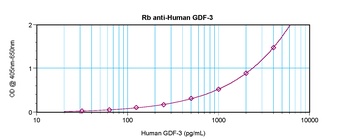
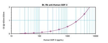
To detect hGDF-3 by sandwich ELISA (using 100 ul/well antibody solution) a concentration of 0.5 - 2.0 ug/ml of this antibody is required. This antigen affinity purified antibody, in conjunction with Biotinylated Anti-Human GDF-3 (orb1272738) as a detection antibody, allows the detection of at least 0.2 - 0.4 ng/well of recombinant hGDF-3.
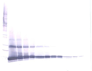
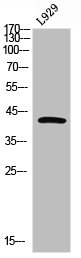
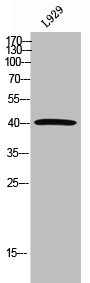
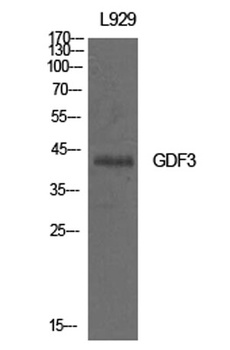
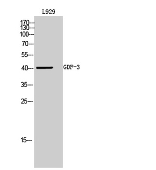
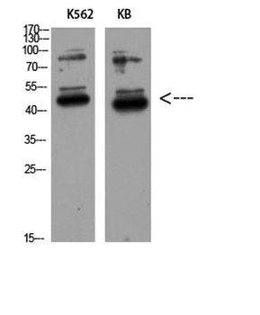
To detect hGDF-3 by Western Blot analysis this antibody can be used at a concentration of 0.1 - 0.2 ug/ml. Used in conjunction with compatible secondary reagents the detection limit for recombinant hGDF-3 is 1.5 - 3.0 ng/lane, under either reducing or non-reducing conditions.
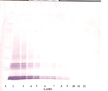
To detect hGDF-3 by Western Blot analysis this antibody can be used at a concentration of 0.1 - 0.2 ug/ml. Used in conjunction with compatible secondary reagents the detection limit for recombinant hGDF-3 is 1.5 - 3.0 ng/lane, under either reducing or non-reducing conditions.
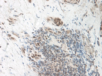
This antibody stained formalin-fixed, paraffin-embedded sections of human breast invasive ductal carcinoma. The recommended concentration is 0.125 ug/ml with an overnight incubation at 4 °C. An HRP-labeled polymer detection system was used with a DAB chromogen. Heat induced antigen retrieval with a pH 6.0 sodium citrate buffer is recommended. Optimal concentrations and conditions may vary. Tissue samples were provided by the Cooperative Human Tissue Network, which is funded by the National Cancer Institute.
- Item 1 of 2
- Item 1 of 3
- Item 1 of 3
- Item 1 of 1
- Item 1 of 2


Submit a review
Filter by Rating

- 5 stars

- 4 stars
- 3 stars
- 2 stars
- 1 stars