You have no items in your shopping cart.
Cart summary
Item 1 of 2
Item 1 of 2
EPHA2 (Phospho-Y588/596) antibody
Catalog Number: orb393275
| Catalog Number | orb393275 |
|---|---|
| Category | Antibodies |
| Description | Rabbit polyclonal antibody to EPHA2 (Phospho-Y588/596) |
| Species/Host | Rabbit |
| Clonality | Polyclonal |
| Tested applications | IF, WB |
| Reactivity | Human, Mouse, Primate, Rat |
| Immunogen | KLH-conjugated synthetic phosphopeptide corresponding to residues surrounding Y588/596 of human EPHA2 protein. The exact sequence is proprietary. |
| Dilution range | WB: 1:500:1000, IF/ICC: 1:100:500 |
| Form/Appearance | Liquid in 0.42% Potassium phosphate, 0.87% Sodium chloride, pH 7.3, 30% glycerol, and 0.01% sodium azide. |
| Conjugation | Unconjugated |
| Target | EPHA2; EPHA3; EPHA4 |
| Entrez | 29210, 2042, 1969, 2043, 13836, 13838 |
| UniProt ID | Q03145, Q03137, P29320, P54764, P29317, O08680, P29319 |
| Source | Rabbit |
| Storage | Shipped at 4°C. Upon delivery aliquot and store at -20°C for one year. Avoid freeze/thaw cycles. |
| Buffer/Preservatives | Liquid in 0.42% Potassium phosphate, 0.87% Sodium chloride, pH 7.3, 30% glycerol, and 0.01% sodium azide. |
| Alternative names | EPHA2; ECK; Ephrin type-A receptor 2; Epithelial c Read more... |
| Note | For research use only |
| Expiration Date | 12 months from date of receipt. |
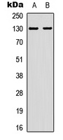
Western blot analysis of EPHA2 (Phospho-Y588/596) expression in A431 (A), mouse brain (B) whole cell lysates. (Predicted band size: 108; 110; 109 kD; Observed band size: 130 kD)
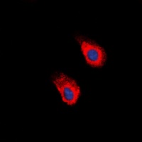
Immunofluorescent analysis of EPHA2 (Phospho-Y588/596) staining in A431 cells. Formalin-fixed cells were permeabilized with 0.1% Triton X-100 in TBS for 5-10 minutes and blocked with 3% BSA-PBS for 30 minutes at room temperature. Cells were probed with the primary antibody in 3% BSA-PBS and incubated overnight at 4 °C in a hidified chamber. Cells were washed with PBST and incubated with a DyLight 594-conjugated secondary antibody (red) in PBS at room temperature in the dark. DAPI was used to stain the cell nuclei (blue).

EPHA2 (Phospho-Y588/596) antibody [orb570660]
ELISA, IF, WB
Human, Mouse, Rat
Rabbit
Polyclonal
Unconjugated
50 μg, 100 μg
Submit a review
Filter by Rating

- 5 stars

- 4 stars
- 3 stars
- 2 stars
- 1 stars