You have no items in your shopping cart.
Cart summary
Item 1 of 7
Item 1 of 7
ENO1 Antibody
Catalog Number: orb654297
| Catalog Number | orb654297 |
|---|---|
| Category | Antibodies |
| Description | ENO1 Antibody |
| Species/Host | Rabbit |
| Clonality | Polyclonal |
| Tested applications | FC, ICC, IF, IHC, WB |
| Reactivity | Human, Monkey, Mouse, Rat |
| Isotype | Rabbit IgG |
| Immunogen | A synthetic peptide corresponding to a sequence of human ENO1 (DDPSRYISPDQLADLYKSFIKD). |
| Concentration | Adding 0.2 ml of distilled water will yield a concentration of 500 μg/ml. |
| Dilution range | Western blot, 0.1-0.25μg/ml, Human, Mouse, Monkey, Rat Immunohistochemistry (Paraffin-embedded Section), 0.5-1μg/ml, Human, Mouse, Rat Immunocytochemistry/Immunofluorescence, 2μg/ml, Human Flow Cytometry, 1-3μg/1x106 cells, Human |
| Form/Appearance | Lyophilized |
| Conjugation | Unconjugated |
| MW | 47 kDa |
| UniProt ID | P06733 |
| Storage | Store at -20˚C for one year from date of receipt. After reconstitution, at 4˚C for one month. It can also be aliquotted and stored frozen at -20˚C for six months. Avoid repeated freeze-thaw cycles. |
| Alternative names | Alpha-enolase; 2-phospho-D-glycerate hydro-lyase; Read more... |
| Note | For research use only |
| Application notes | Tested Species: In-house tested species with positive results. Other applications have not been tested. Optimal dilutions should be determined by end users. Add 0.2ml of distilled water will yield a concentration of 500ug/ml. |
| Expiration Date | 12 months from date of receipt. |
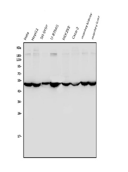
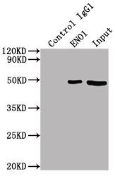
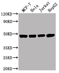
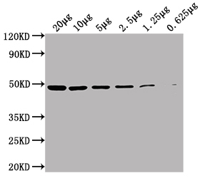
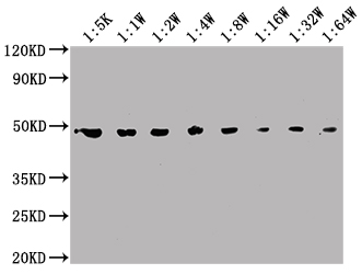
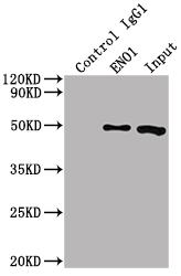
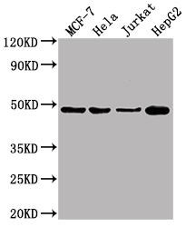
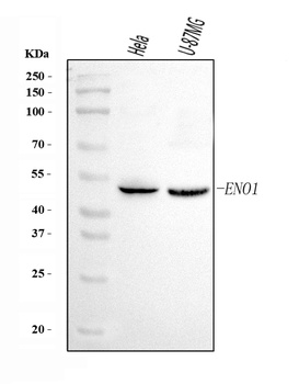
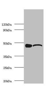
Western blot analysis of ENO1 using anti-ENO1 antibody (orb654297). Electrophoresis was performed on a 5-20% SDS-PAGE gel at 70V (Stacking gel)/90V (Resolving gel) for 2-3 hours. The sample well of each lane was loaded with 50ug of sample under reducing conditions. Lane 1: human HeLa whole cell lysates, Lane 2: human HepG2 whole cell lysates, Lane 3: human SH-SY5Y whole cell lysates, Lane 4: human U-87MG whole cell lysates, Lane 5: human HEK293 whole cell lysates, Lane 6: human Caco-2 whole cell lysates, Lane 7: monkey kidney tissue lysates, Lane 8: monkey liver tissue lysates. After Electrophoresis, proteins were transferred to a Nitrocellulose membrane at 150mA for 50-90 minutes. Blocked the membrane with 5% Non-fat Milk/TBS for 1.5 hour at RT. The membrane was incubated with rabbit anti-ENO1 antigen affinity purified polyclonal antibody (Catalog # orb654297) at 0.25 μg/mL overnight at 4°C, then washed with TBS-0.1%Tween 3 times with 5 minutes each and probed with a goat anti-rabbit IgG-HRP secondary antibody at a dilution of 1:10000 for 1.5 hour at RT. The signal is developed using an Enhanced Chemiluminescent detection (ECL) kit (Catalog # orb90503) with Tanon 5200 system. A specific band was detected for ENO1 at approximately 47KD. The expected band size for ENO1 is at 47KD.
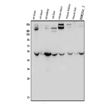
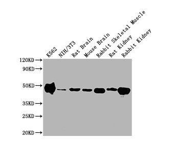
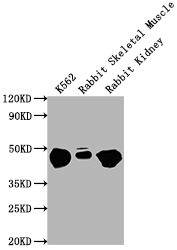
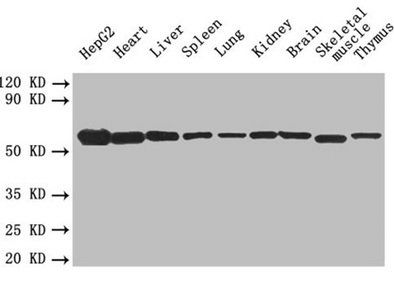
Western blot analysis of ENO1 using anti-ENO1 antibody (orb654297). Electrophoresis was performed on a 5-20% SDS-PAGE gel at 70V (Stacking gel)/90V (Resolving gel) for 2-3 hours. The sample well of each lane was loaded with 50ug of sample under reducing conditions. Lane 1: rat brain tissue lysates, Lane 2: rat heart tissue lysates, Lane 3: rat kidney tissue lysates, Lane 4: rat liver tissue lysates, Lane 5: mouse brain tissue lysates, Lane 6: mouse kidney tissue lysates, Lane 7: mouse liver tissue lysates, Lane 8: mouse RAW264.7 whole cell lysates. After Electrophoresis, proteins were transferred to a Nitrocellulose membrane at 150mA for 50-90 minutes. Blocked the membrane with 5% Non-fat Milk/TBS for 1.5 hour at RT. The membrane was incubated with rabbit anti-ENO1 antigen affinity purified polyclonal antibody (Catalog # orb654297) at 0.25 μg/mL overnight at 4°C, then washed with TBS-0.1%Tween 3 times with 5 minutes each and probed with a goat anti-rabbit IgG-HRP secondary antibody at a dilution of 1:10000 for 1.5 hour at RT. The signal is developed using an Enhanced Chemiluminescent detection (ECL) kit (Catalog # orb90503) with Tanon 5200 system. A specific band was detected for ENO1 at approximately 47KD. The expected band size for ENO1 is at 47KD.
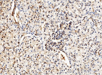
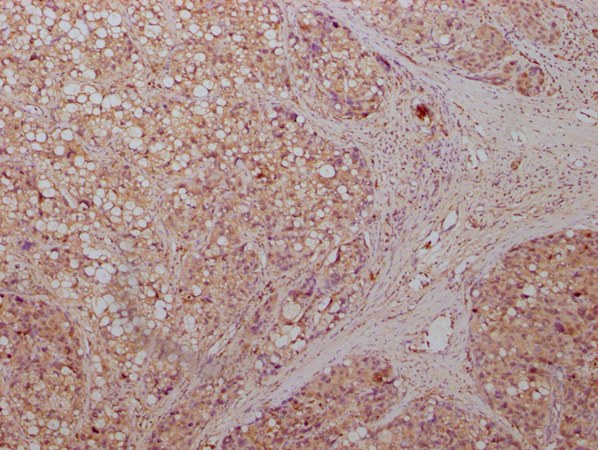
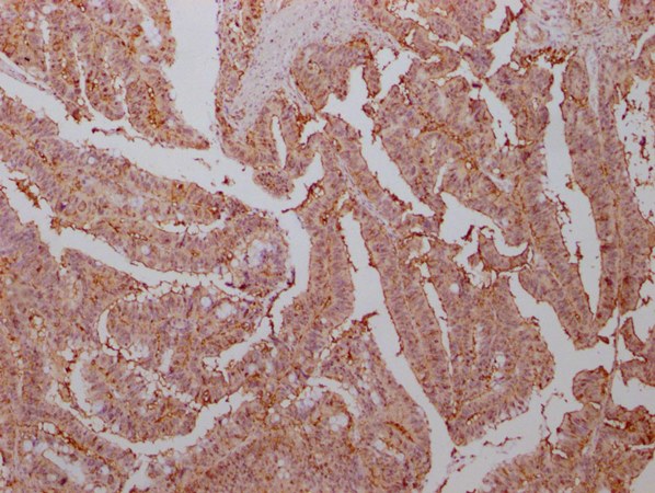
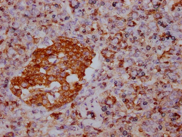
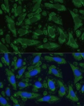
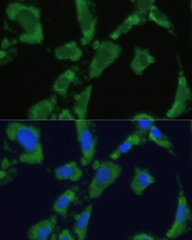
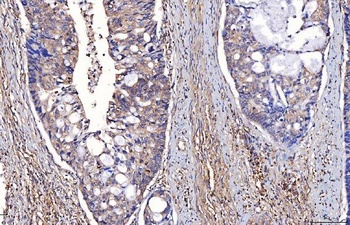
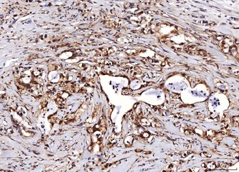
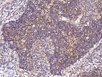
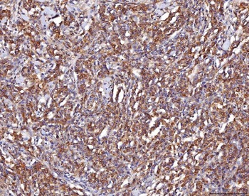
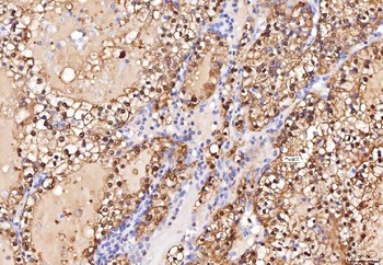
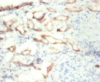
IHC analysis of ENO1 using anti-ENO1 antibody (orb654297). ENO1 was detected in paraffin-embedded section of human liver cancer tissue. Heat mediated antigen retrieval was performed in EDTA buffer (pH8.0, epitope retrieval solution). The tissue section was blocked with 10% goat serum. The tissue section was then incubated with 1μg/ml rabbit anti-ENO1 Antibody (orb654297) overnight at 4°C. Biotinylated goat anti-rabbit IgG was used as secondary antibody and incubated for 30 minutes at 37°C. The tissue section was developed using Strepavidin-Biotin-Complex (SABC) (Catalog # orb90444) with DAB as the chromogen.
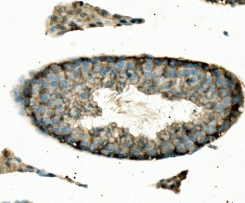
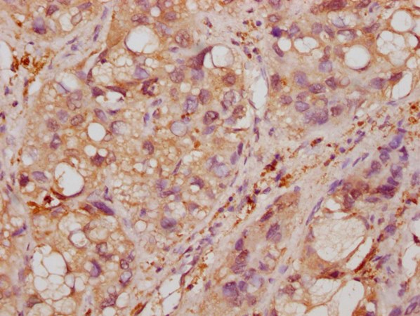
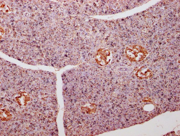
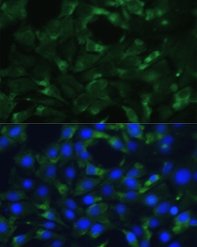
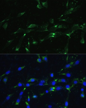
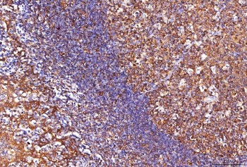
IHC analysis of ENO1 using anti-ENO1 antibody (orb654297). ENO1 was detected in paraffin-embedded section of mouse testis tissue. Heat mediated antigen retrieval was performed in EDTA buffer (pH8.0, epitope retrieval solution). The tissue section was blocked with 10% goat serum. The tissue section was then incubated with 1μg/ml rabbit anti-ENO1 Antibody (orb654297) overnight at 4°C. Biotinylated goat anti-rabbit IgG was used as secondary antibody and incubated for 30 minutes at 37°C. The tissue section was developed using Strepavidin-Biotin-Complex (SABC) (Catalog # orb90444) with DAB as the chromogen.
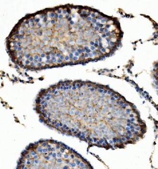
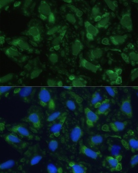
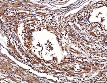
IHC analysis of ENO1 using anti-ENO1 antibody (orb654297). ENO1 was detected in paraffin-embedded section of rat testis tissue. Heat mediated antigen retrieval was performed in EDTA buffer (pH8.0, epitope retrieval solution). The tissue section was blocked with 10% goat serum. The tissue section was then incubated with 1μg/ml rabbit anti-ENO1 Antibody (orb654297) overnight at 4°C. Biotinylated goat anti-rabbit IgG was used as secondary antibody and incubated for 30 minutes at 37°C. The tissue section was developed using Strepavidin-Biotin-Complex (SABC) (Catalog # orb90444) with DAB as the chromogen.
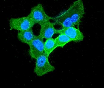
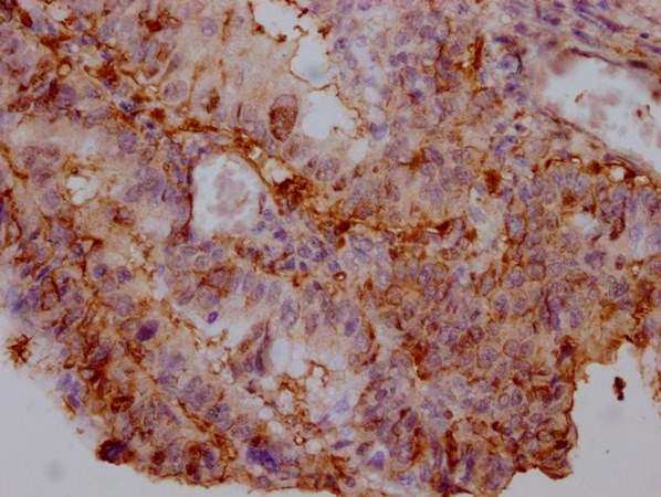
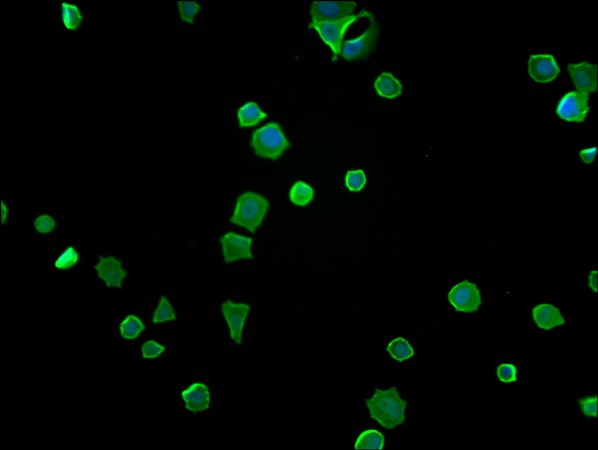
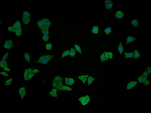
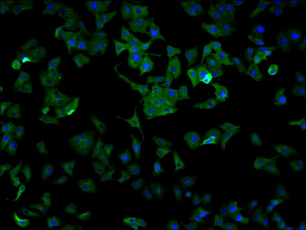
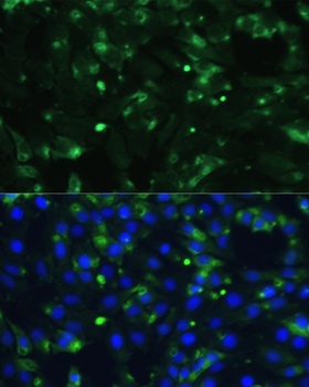
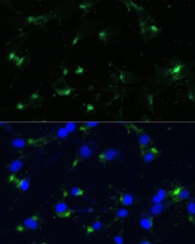
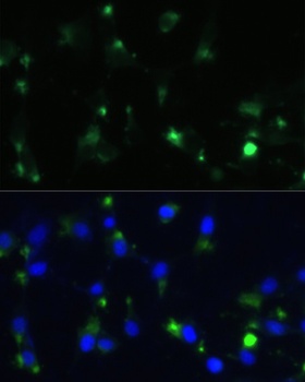
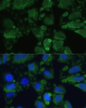
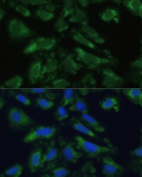
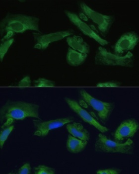
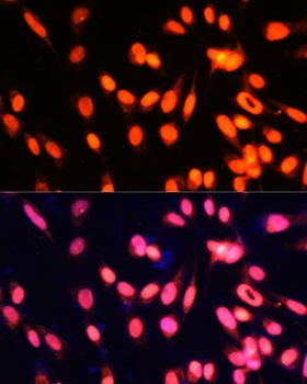
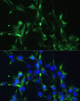
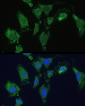
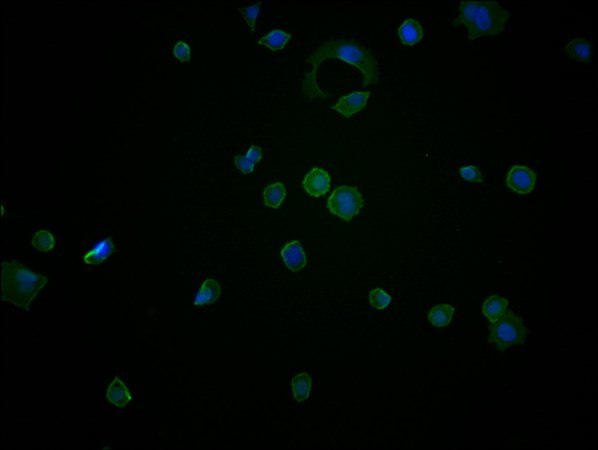
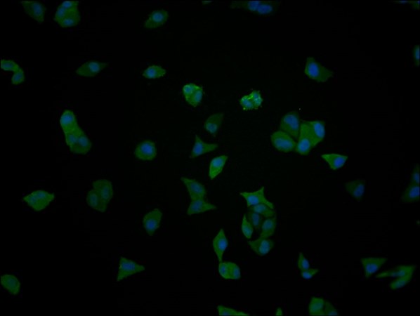
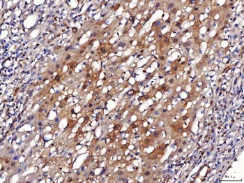
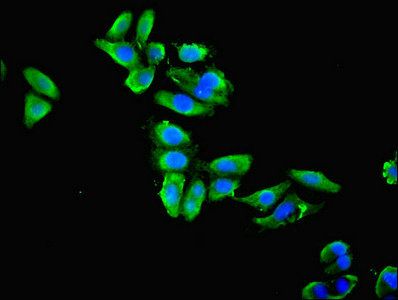
IF analysis of ENO1 using anti-ENO1 antibody (orb654297). ENO1 was detected in immunocytochemical section of A431 cells. Enzyme antigen retrieval was performed using IHC enzyme antigen retrieval reagent (orb90553) for 15 mins. The cells were blocked with 10% goat serum. And then incubated with 2μg/mL rabbit anti-ENO1 Antibody (orb654297) overnight at 4°C. DyLight®488 Conjugated Goat Anti-Rabbit IgG was used as secondary antibody at 1:100 dilution and incubated for 30 minutes at 37°C. The section was counterstained with DAPI. Visualize using a fluorescence microscope and filter sets appropriate for the label used.
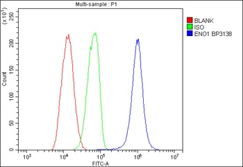
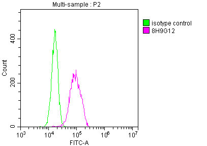
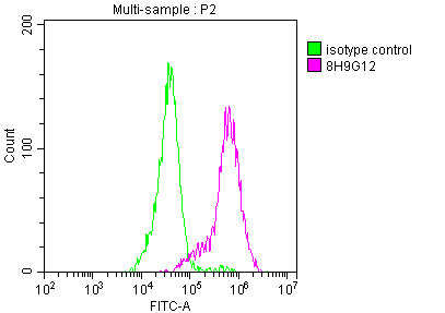
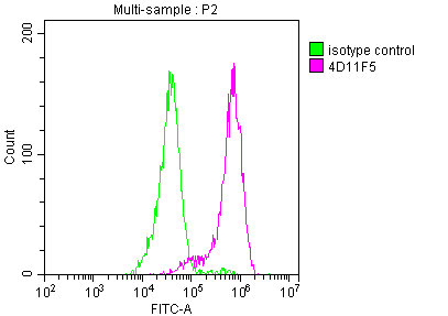
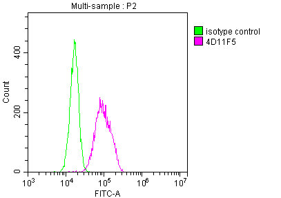
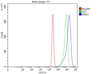
Flow Cytometry analysis of HL-60 cells using anti-ENO1 antibody (orb654297). Overlay histogram showing HL-60 cells stained with orb654297 (Blue line). The cells were blocked with 10% normal goat serum. And then incubated with rabbit anti-ENO1 Antibody (orb654297, 1μg/1x10^6 cells) for 30 min at 20°C. DyLight®488 conjugated goat anti-rabbit IgG (5-10μg/1x10^6 cells) was used as secondary antibody for 30 minutes at 20°C. Isotype control antibody (Green line) was rabbit IgG (1μg/1x10^6) used under the same conditions. Unlabelled sample (Red line) was also used as a control.
- Item 1 of 16
ENO1 antibody [orb688875]
ELISA, FC, IF, IHC, IP, WB
Human, Mouse, Rabbit, Rat
Mouse
Monoclonal
Unconjugated
100 μl, 50 μl - Item 1 of 15
- Item 1 of 9
ENO1 antibody [orb688876]
ELISA, FC, IF, IP, WB
Human, Mouse, Rabbit, Rat
Mouse
Monoclonal
Unconjugated
100 μl, 50 μl - Item 1 of 10
- Item 1 of 4
Alpha-enolase antibody [orb239776]
ELISA, IF, IHC, WB
Human, Mouse
Rabbit
Polyclonal
Unconjugated
100 μg, 50 μg
Submit a review
Filter by Rating

- 5 stars

- 4 stars
- 3 stars
- 2 stars
- 1 stars