You have no items in your shopping cart.
Cart summary
Item 1 of 4
Item 1 of 4
ENO1 Antibody
Catalog Number: orb1263818

| Catalog Number | orb1263818 |
|---|---|
| Category | Antibodies |
| Description | ENO1 Antibody |
| Target | ENO1 |
| Clonality | Polyclonal |
| Isotype | Rabbit Ig |
| Conjugation | Unconjugated |
| Reactivity | Human, Mouse |
| Predicted Reactivity | Bovine, Gallus, Monkey, Rat, Xenopus |
| Form/Appearance | Liquid |
| Concentration | batch dependent |
| Buffer/Preservatives | Supplied in PBS with 0.09% (W/V) sodium azide. |
| Immunogen | This ENO1 antibody is generated from rabbits immunized with a KLH conjugated synthetic peptide between 178-205 amino acids from the Central region of human ENO1. |
| UniProt ID | P06733 |
| MW | 47 kDa |
| Tested applications | FC, IF, IHC-P, WB |
| Application notes | For WB starting dilution is: 1:1000For IHC-P starting dilution is: 1:10~50For FACS starting dilution is: 1:10~50For IF starting dilution is: 1:10~50 |
| Antibody Type | Primary Antibody |
| Storage | Maintain refrigerated at 2-8°C for up to 2 weeks. For long term storage store at -20°C in small aliquots to prevent freeze-thaw cycles. |
| Alternative names | Alpha-enolase, 2-phospho-D-glycerate hydro-lyase, Read more... |
| Note | For research use only |
| NCBI | P06733 |
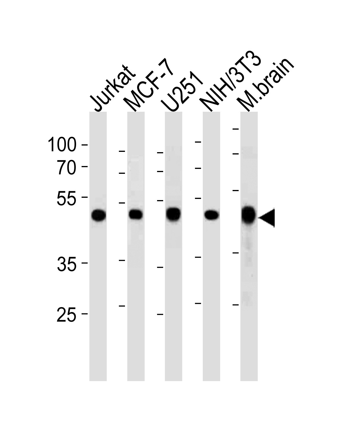
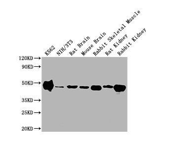
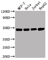
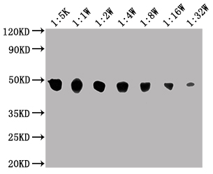
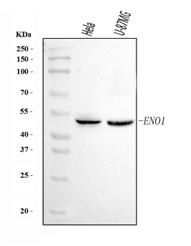
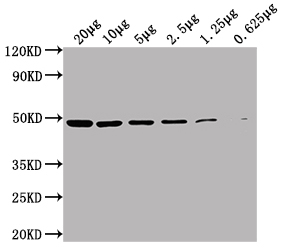
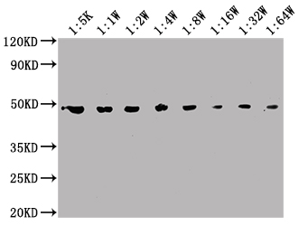
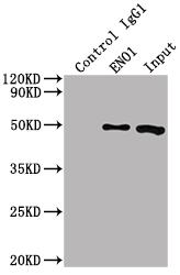
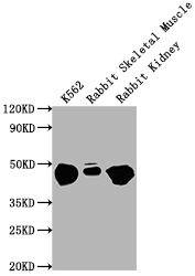
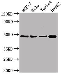
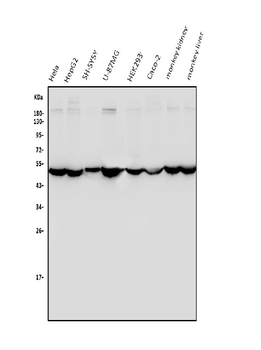
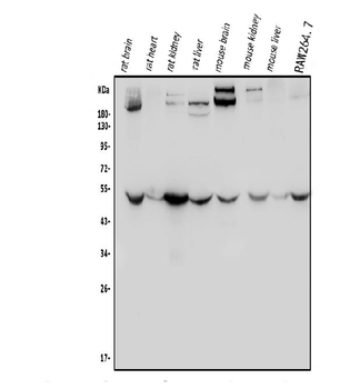
Western blot analysis in Jurkat, MCF-7, U251, mouse NIH/3T3 cell line and mouse brain tissue lysates (35 ug/lane).
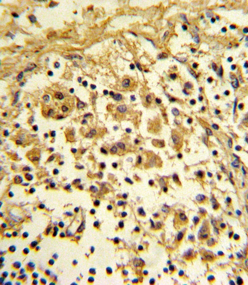
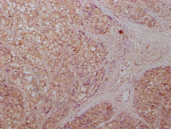
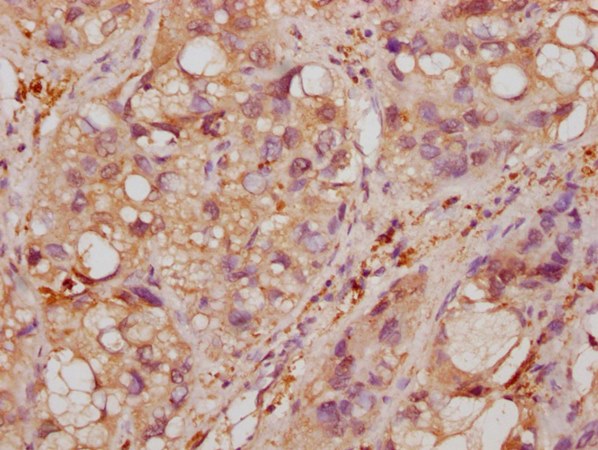
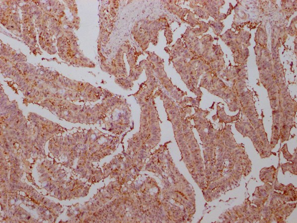
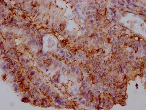
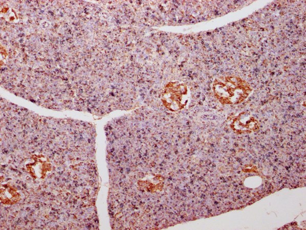
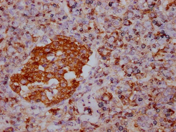
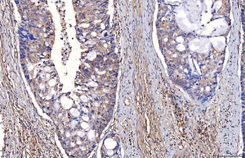
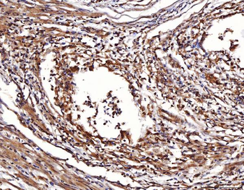
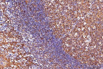
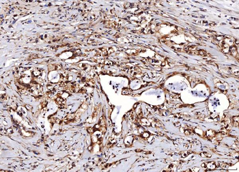
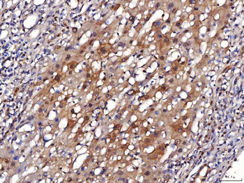
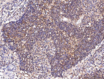
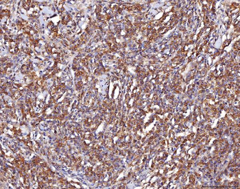
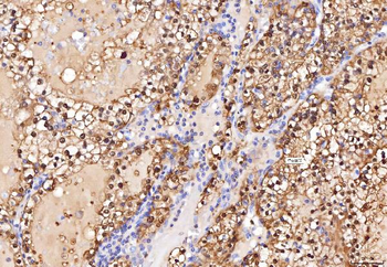
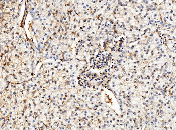
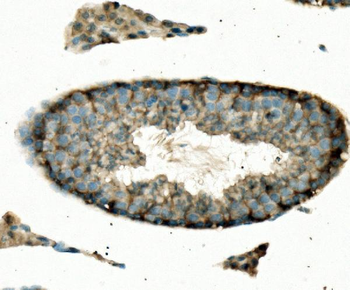
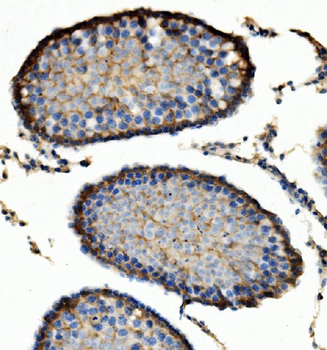
Formalin-fixed and paraffin-embedded human lymph reacted with ENO1 Antibody, which was peroxidase-conjugated to the secondary antibody, followed by DAB staining.
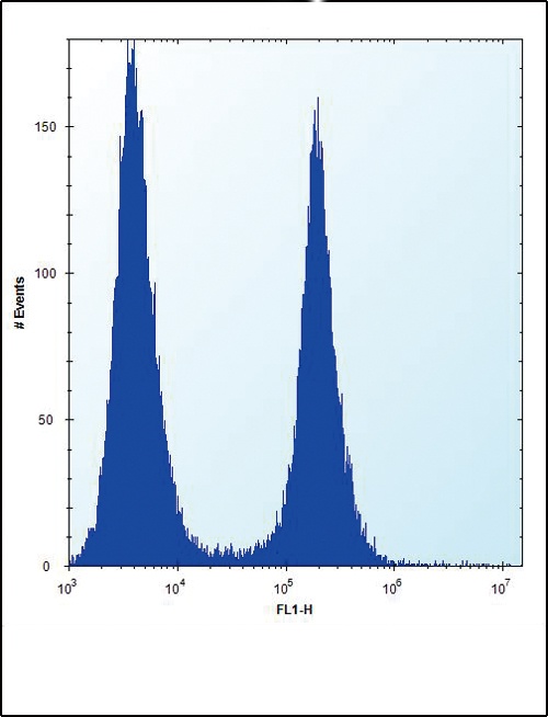
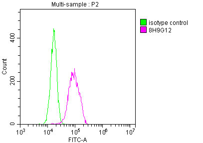
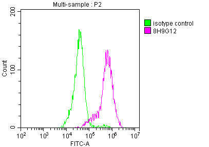
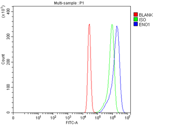
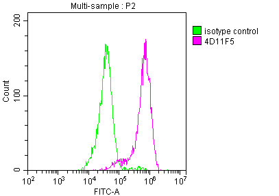
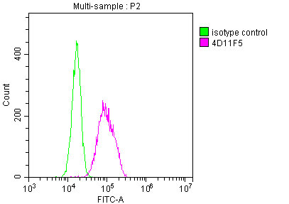
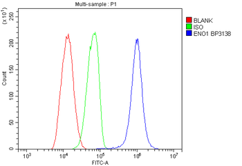
Flow cytometric analysis of Hela cells (right histogram) compared to a negative control cell (left histogram). FITC-conjugated donkey-anti-rabbit secondary antibodies were used for the analysis.
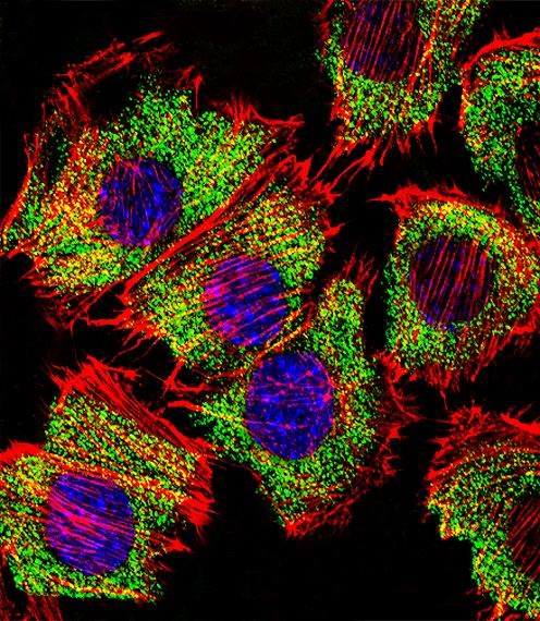
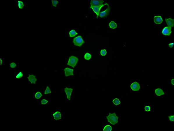
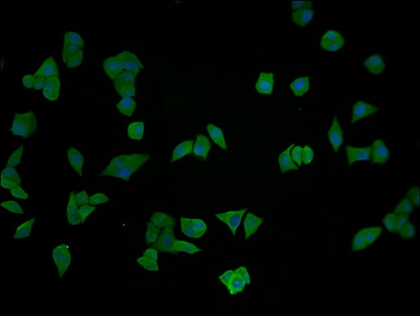
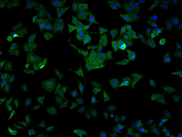
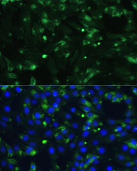
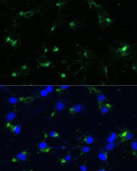
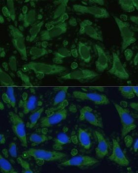
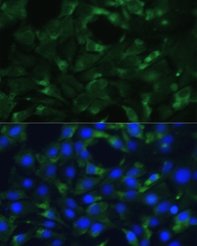
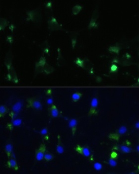
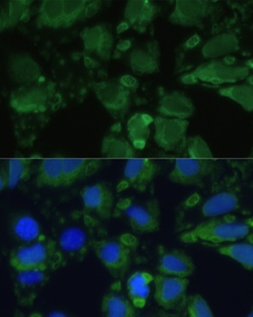
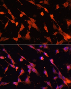
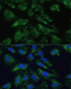
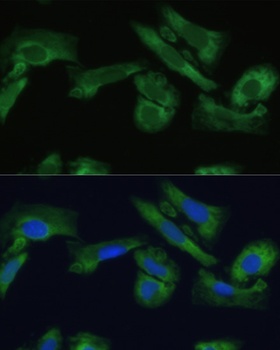
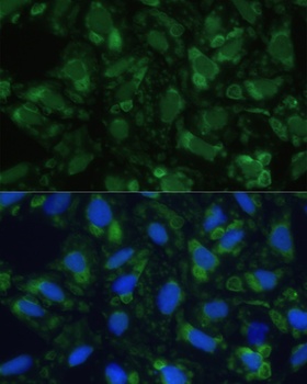
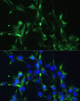
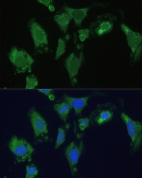
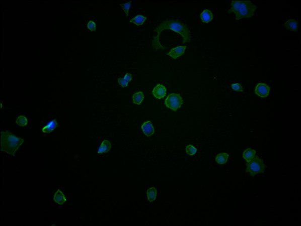
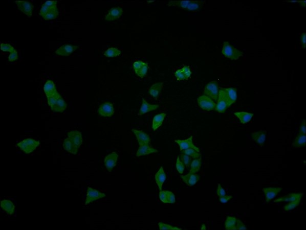
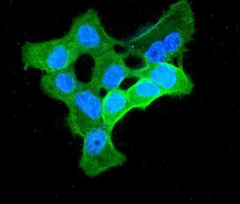
Fluorescent confocal image of C2C12 cell stained with ENO1 Antibody.C2C12 cells were fixed with 4% PFA (20 min), permeabilized with Triton X-100 (0.1%, 10 min), then incubated with ENO1 primary antibody (1:25). For secondary antibody, Alexa Fluor 488 conjugated donkey anti-rabbit antibody (green) was used (1:400).Cytoplasmic actin was counterstained with Alexa Fluor 555 (red) conjugated Phalloidin (7 units/ml). Nuclei were counterstained with DAPI (blue) (10 ug/ml, 10 min). ENO1 immunoreactivity is localized to Cytoplasm significantly.
- Item 1 of 16
ENO1 Monoclonal Antibody [orb688875]
ELISA, FC, IF, IHC, IP, WB
Human, Mouse, Rabbit, Rat
Mouse
Monoclonal
Unconjugated
50 μl, 100 μl - Item 1 of 15
- Item 1 of 10
- Item 1 of 9
ENO1 Monoclonal Antibody [orb688876]
ELISA, FC, IF, IP, WB
Human, Mouse, Rabbit, Rat
Mouse
Monoclonal
Unconjugated
100 μl, 50 μl - Item 1 of 7
Anti-ENO1 Antibody [orb654297]
FC, ICC, IF, IHC, WB
Human, Monkey, Mouse, Rat
Rabbit
Polyclonal
Unconjugated
100 μg, 10 μg