You have no items in your shopping cart.
Cart summary
Item 1 of 11
Item 1 of 11
EHD2 antibody
Catalog Number: orb18895
| Catalog Number | orb18895 |
|---|---|
| Category | Antibodies |
| Description | Goat polyclonal antibody to EHD2 |
| Species/Host | Goat |
| Clonality | Polyclonal |
| Tested applications | ELISA, FC, IF, WB |
| Reactivity | Bovine, Canine, Human, Mouse, Rat |
| Dilution range | ELISA: 1:64000, WB: 0.1-0.3 μg/ml, IHC-P: 10ug/ml |
| Conjugation | Unconjugated |
| MW | 61.2 |
| Target | EHD2 |
| Entrez | 30846 |
| Protein Sequence | CRLVPPSKRRHKGSA |
| Storage | Aliquot and store at -20°C. Minimize freezing and thawing. |
| Buffer/Preservatives | Supplied at 0.5 mg/ml in Tris saline, 0.02% sodium azide, pH 7.3 with 0.5% bovine serum albumin. Aliquot and store at -20°C. Minimize freezing and thawing. |
| Alternative names | anti EHD2 antibody, anti PAST2 antibody, anti EH-d Read more... |
| Note | For research use only |
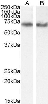
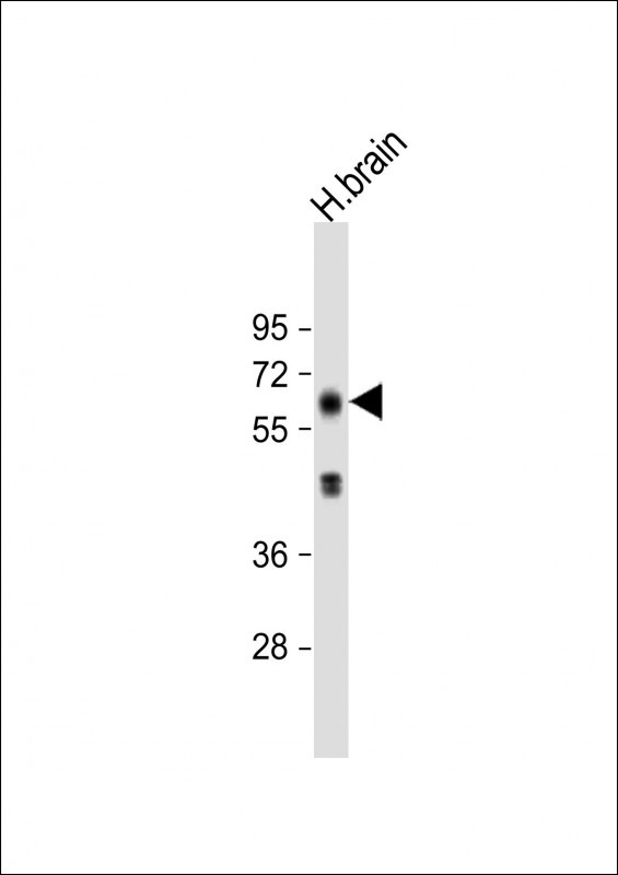
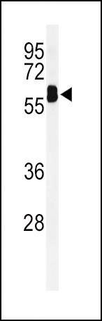
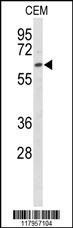
| Application notes | ELISA: Peptide ELISA: antibody detection limit dilution 1:32000.WB: Approx 60kDa band observed in Human Heart lysates (calculated MW of 61.2kDa according to NP_055416.2). In transfected HEK293 transiently expressing EHD2 a band of approx. 62kDa is observed. This band is not observed in the non-transfected HEK293. Recommended concentration: 0.003-0.01 μg/ml. |
| Expiration Date | 12 months from date of receipt. |
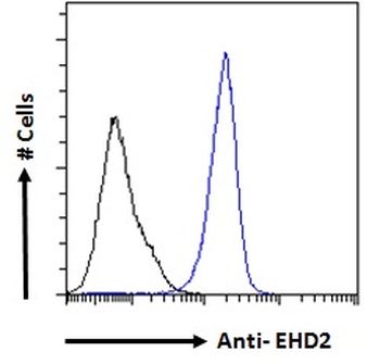
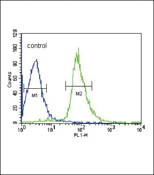
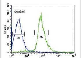
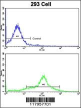
orb18895 Flow cytometric analysis of paraformaldehyde fixed A431 cells (blue line), permeabilized with 0.5% Triton. Primary incubation 1hr (10ug/ml) followed by Alexa Fluor 488 secondary antibody (1ug/ml). IgG control: Unimmunized goat IgG (black line) followed by Alexa Fluor 488 secondary antibody.
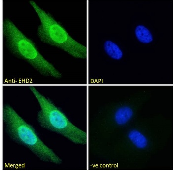
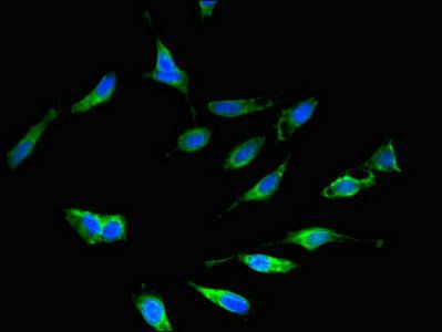
orb18895 Immunofluorescence analysis of paraformaldehyde fixed HeLa cells, permeabilized with 0.15% Triton. Primary incubation 1hr (10ug/ml) followed by Alexa Fluor 488 secondary antibody (2ug/ml), showing nuclear and cytoplasmic staining. The nuclear stain is DAPI (blue). Negative control: Unimmunized goat IgG (10ug/ml) followed by Alexa Fluor 488 secondary antibody (2ug/ml).
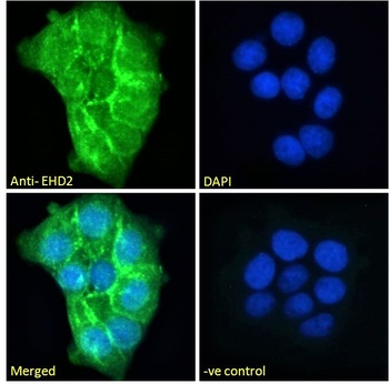
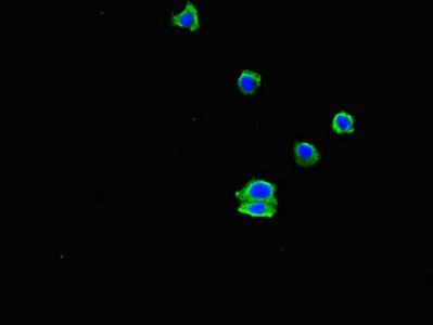
orb18895 Immunofluorescence analysis of paraformaldehyde fixed A431 cells, permeabilized with 0.15% Triton. Primary incubation 1hr (10ug/ml) followed by Alexa Fluor 488 secondary antibody (2ug/ml), showing plasma membrane staining. The nuclear stain is DAPI (blue). Negative control: Unimmunized goat IgG (10ug/ml) followed by Alexa Fluor 488 secondary antibody (2ug/ml).
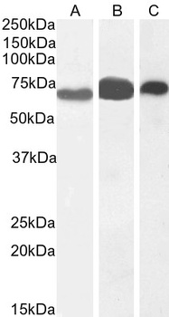
orb18895 (0.1g/ml) staining of Human Lung (A), Colon (B) and Placenta (C) lysate (35µg protein in RIPA buffer). Detected by chemiluminescence.
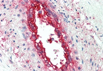
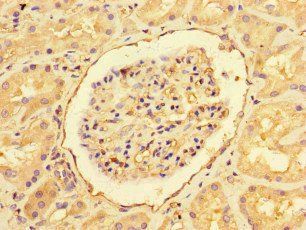
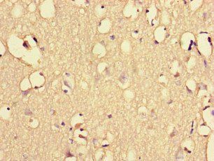
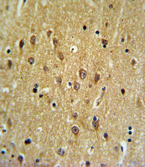
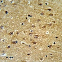
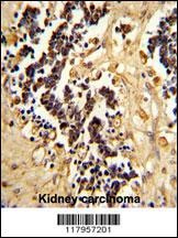
orb18895 (5µg/ml) staining of paraffin embedded Human Vessel. Steamed antigen retrieval with citrate buffer pH 6, AP-staining. Data obtained from a previous batch, not on sale.
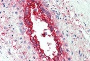
Immunohistochemical staining of Human Vessel using EHD2 antibody
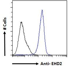
Flow Cytometry analysis of A431 cells of EHD2 antibody

Immunohistochemical staining of HeLa cells of EHD2 antibody

Immunohistochemical staining of A431 cells of EHD2 antibody

Western blot analysis of Human Lung lysate using EHD2 antibody
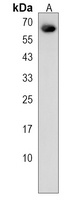
Western blot analysis of A549 (Lane 1) and (0.3µg/ml) HeLa (Lane 2) cell lysate using paroduct name
- Item 1 of 3
- Item 1 of 4
- Item 1 of 3
- Item 1 of 2
- Item 1 of 3
Submit a review
Filter by Rating

- 5 stars

- 4 stars
- 3 stars
- 2 stars
- 1 stars