You have no items in your shopping cart.
Cart summary
Item 1 of 3
Item 1 of 3
EAAC1 Antibody: APC
Catalog Number: orb149928

| Catalog Number | orb149928 |
|---|---|
| Category | Antibodies |
| Description | Mouse monoclonal antibody against SLC1A1 conjugated to APC |
| Species/Host | Mouse |
| Clonality | Monoclonal |
| Clone Number | N180/41 (Formerly sold as S180-41) |
| Tested applications | ICC, IF, IHC |
| Reactivity | Human, Rat |
| Isotype | IgG1 |
| Immunogen | Fusion protein amino acids 1-524 (full length) of human EAAC1 |
| Concentration | 1 mg/ml |
| Dilution range | WB (1:1000), ICC/IF (1:100);optimal dilutions for assays should be determined by the user. |
| Conjugation | APC |
| MW | 60kDa |
| Target | EAAT3 |
| Entrez | 6505 |
| UniProt ID | P43005 |
| NCBI | NP_004161.4 |
| Storage | Conjugated antibodies should be stored according to the product label |
| Buffer/Preservatives | 95.64mM Phosphate, 2.48mM MES and 2mM EDTA |
| Alternative names | Excitatory amino-acid carrier 1 antibody, excitato Read more... |
| Note | For research use only |
| Application notes | 5 µg of SMC-406 was sufficient for immunoprecipitation of EAAC1 from 50 µg of rat brain lysate. |
| Expiration Date | 12 months from date of receipt. |

Immunocytochemistry/Immunofluorescence analysis using Mouse Anti-EAAC1 Monoclonal Antibody, Clone N180/41. Tissue: Neuroblastoma cells (SH-SY5Y). Species: Human. Fixation: 4% PFA for 15 min. Primary Antibody: Mouse Anti-EAAC1 Monoclonal Antibody at 1:100 for overnight at 4°C with slow rocking. Secondary Antibody: AlexaFluor 488 at 1:1000 for 1 hour at RT. Counterstain: Phalloidin-iFluor 647 (red) F-Actin stain; Hoechst (blue) nuclear stain at 1:800, 1.6mM for 20 min at RT. (A) Hoechst (blue) nuclear stain. (B) Phalloidin-iFluor 647 (red) F-Actin stain. (C) EAAC1 Antibody (D) Composite.
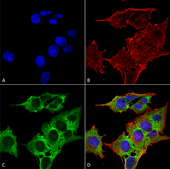
Immunocytochemistry/Immunofluorescence analysis using Mouse Anti-EAAC1 Monoclonal Antibody, Clone N180/41. Tissue: Neuroblastoma cell line (SK-N-BE). Species: Human. Fixation: 4% Formaldehyde for 15 min at RT. Primary Antibody: Mouse Anti-EAAC1 Monoclonal Antibody at 1:100 for 60 min at RT. Secondary Antibody: Goat Anti-Mouse ATTO 488 at 1:200 for 60 min at RT. Counterstain: Phalloidin Texas Red F-Actin stain; DAPI (blue) nuclear stain at 1:1000, 1:5000 for 60 min at RT, 5 min at RT. Localization: Cytoplasm, Membrane. Magnification: 60X. (A) DAPI (blue) nuclear stain. (B) Phalloidin Texas Red F-Actin stain. (C) EAAC1 Antibody. (D) Composite.
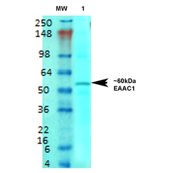
Western Blot analysis of Rat brain membrane lysate showing detection of EAAT3 protein using Mouse Anti-EAAT3 Monoclonal Antibody, Clone N180/41. Primary Antibody: Mouse Anti-EAAT3 Monoclonal Antibody at 1:1000.
