You have no items in your shopping cart.
Cart summary
Item 1 of 7
Item 1 of 7
DVL1 Antibody
Catalog Number: orb1269676

| Catalog Number | orb1269676 |
|---|---|
| Category | Antibodies |
| Description | DVL1 Antibody |
| Species/Host | Rabbit |
| Clonality | Polyclonal |
| Tested applications | IF, IHC-P, WB |
| Predicted Reactivity | Rat |
| Reactivity | Human, Mouse |
| Isotype | Rabbit Ig |
| Immunogen | This DVL1 antibody is generated from rabbits immunized with a KLH conjugated synthetic peptide between 442-470 amino acids from the Central region of human DVL1. |
| Antibody Type | Primary Antibody |
| Concentration | batch dependent |
| Form/Appearance | Liquid |
| Conjugation | Unconjugated |
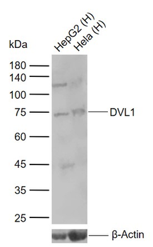
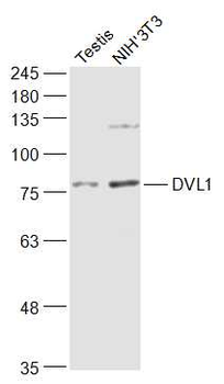
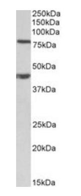
| MW | 75 kDa |
| Target | DVL1 |
| UniProt ID | O14640 |
| NCBI | O14640 |
| Storage | Maintain refrigerated at 2-8°C for up to 2 weeks. For long term storage store at -20°C in small aliquots to prevent freeze-thaw cycles. |
| Buffer/Preservatives | Supplied in PBS with 0.09% (W/V) sodium azide. |
| Alternative names | Segment polarity protein dishevelled homolog DVL-1 Read more... |
| Note | For research use only |
| Application notes | For IF starting dilution is: 1:25For IHC-P starting dilution is: 1:25For WB starting dilution is: 1:1000 |
| Expiration Date | 12 months from date of receipt. |

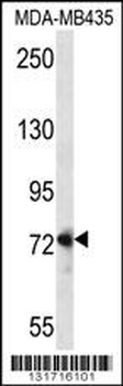
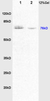
Western blot analysis in MDA-MB435 cell line lysates (35 ug/lane).
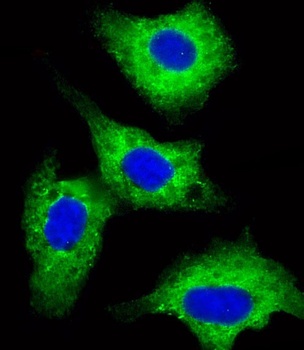
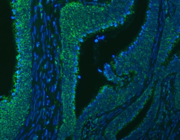
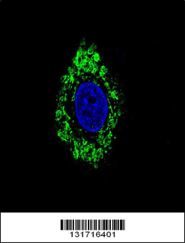
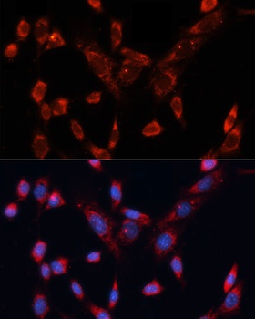
Immunofluorescent analysis of 4% paraformaldehyde-fixed, 0. 1% Triton X-100 permeabilized HepG2 (human liver hepatocellular carcinoma cell line) cells labeling Pdx1 with antibody at 1/25 dilution, followed by Dylight 488-conjugated goat anti-rabbit IgG (NK179883) secondary antibody at 1/200 dilution (green). Immunofluorescence image showing cytoplasm staining on HepG2 cell line. The nuclear counter stain is DAPI (blue).
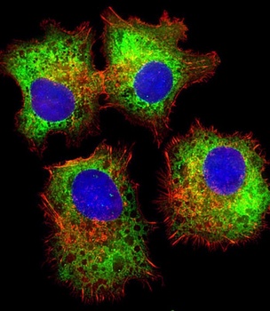
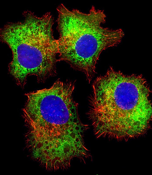
Immunofluorescent analysis of 4% paraformaldehyde-fixed, 0. 1% Triton X-100 permeabilized HepG2 (human liver hepatocellular carcinoma cell line) cells labeling Pdx1 with antibody at 1/25 dilution, followed by Dylight 488-conjugated goat anti-rabbit IgG (NK179883) secondary antibody at 1/200 dilution (green). Immunofluorescence image showing cytoplasm staining on HepG2 cell line. Cytoplasmic actin is detected with Dylight 554 Phalloidin (PD18466410) at 1/100 dilution (red). The nuclear counter stain is DAPI (blue).

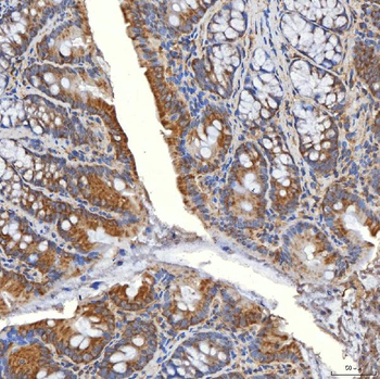
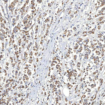
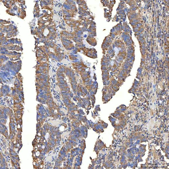
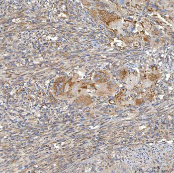
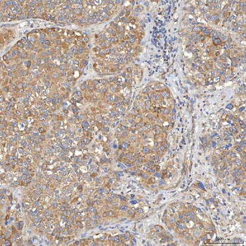
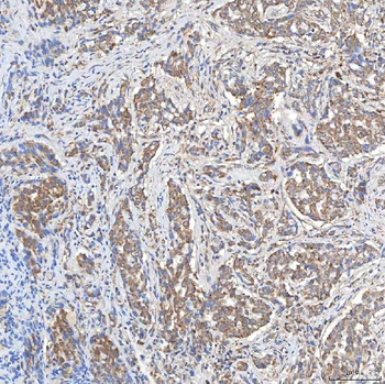
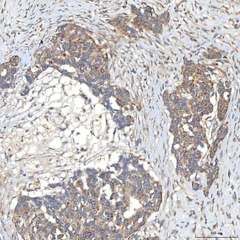
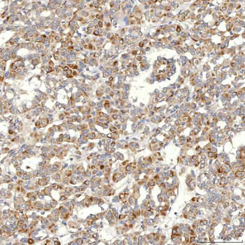
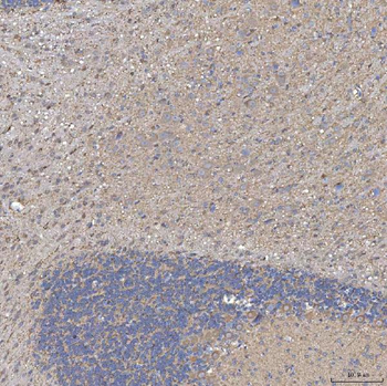
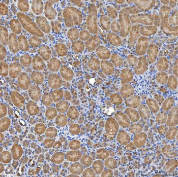
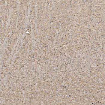
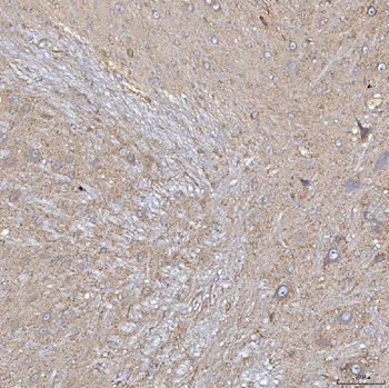
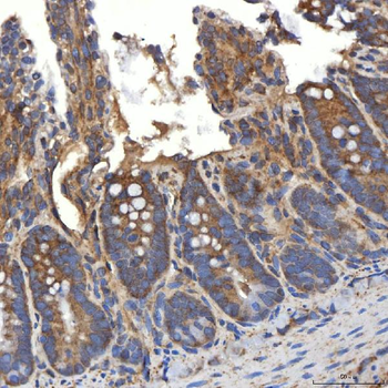
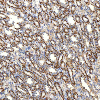
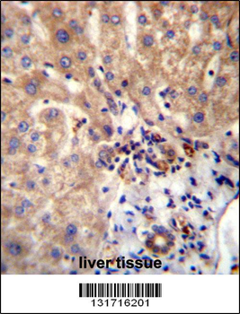
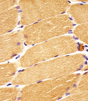
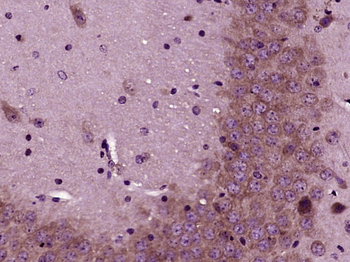
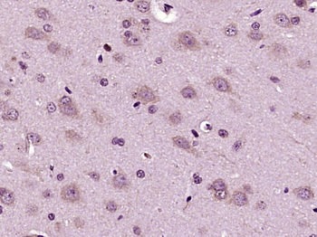
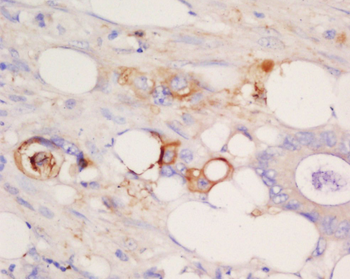
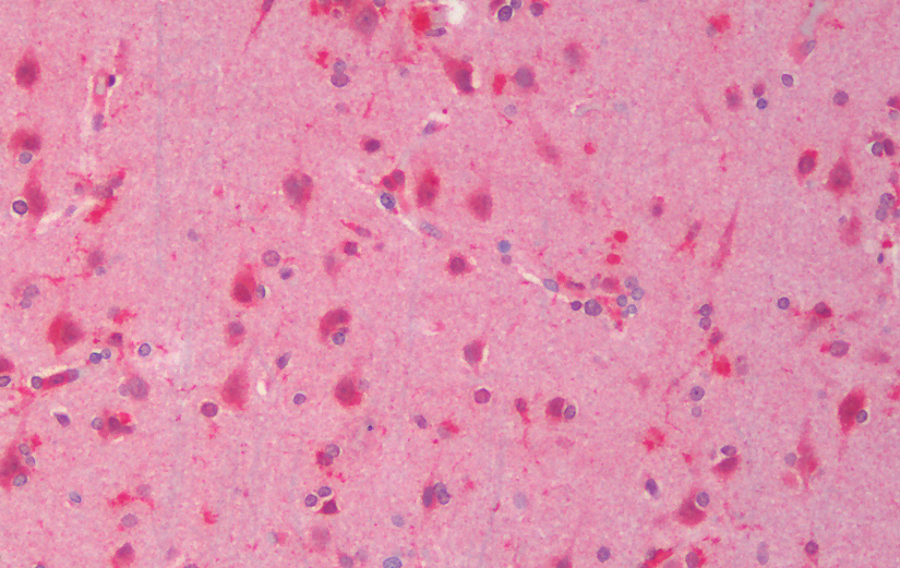
Antibody staining DVL1 in human skeletal muscle sections by Immunohistochemistry (IHC-P - paraformaldehyde-fixed, paraffin-embedded sections).
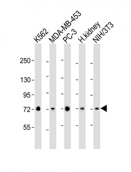
Western Blot at 1:2000 dilution Lane 1: K562 whole cell lysate Lane 2: MDA-MB-453 whole cell lysate Lane 3: PC-3 whole cell lysate Lane 4: human kidney lysate Lane 5: NIH/3T3 whole cell lysate Lysates/proteins at 20 ug per lane.
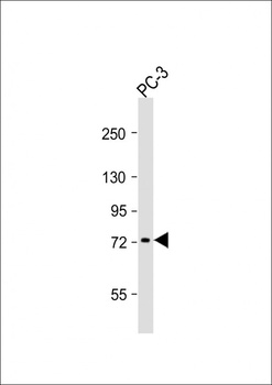
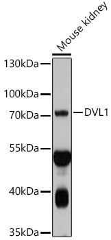
Western Blot at 1:1000 dilution + PC-3 whole cell lysates Lysates/proteins at 20 ug per lane.
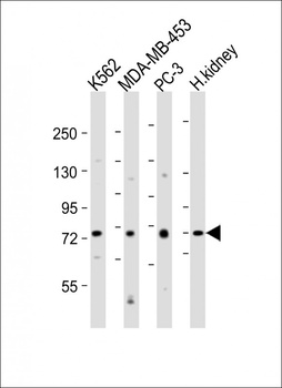
Western Blot at 1:2000 dilution Lane 1: K562 whole cell lysates Lane 2: MDA-MB-453 whole cell lysates Lane 3: PC-3 whole cell lysates Lane 4: human kidney lysates Lysates/proteins at 20 ug per lane.
- Item 1 of 16
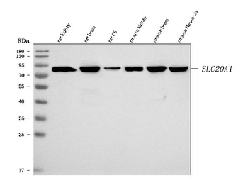
Anti-SLC20A1 Antibody [orb1474852]
ELISA, IF, IHC, WB
Human, Mouse, Rat
Rabbit
Polyclonal
Unconjugated
10 μg, 100 μg - Item 1 of 7
DVL1 Antibody (Center) [orb1937565]
IF, IHC-P, WB
Rat
Human, Mouse
Rabbit
Polyclonal
Unconjugated
100 μl, 50 μl - Item 1 of 6
DVL1 Rabbit Polyclonal Antibody [orb10561]
IF, IHC-Fr, IHC-P, WB
Mouse, Rat
Human, Mouse, Rat
Rabbit
Polyclonal
Unconjugated
200 μl, 50 μl, 100 μl - Item 1 of 2
Goat anti-Dvl1 (mouse) Antibody [orb99056]
ELISA, IHC, WB
Bovine, Human, Mouse, Porcine, Rat
Goat
Polyclonal
Unconjugated
100 μg - Item 1 of 4