You have no items in your shopping cart.
Cart summary
Item 1 of 4
Item 1 of 4
DSTN Antibody (Center)
Catalog Number: orb1928055

| Catalog Number | orb1928055 |
|---|---|
| Category | Antibodies |
| Description | Affinity Purified Rabbit Polyclonal Antibody (Pab) |
| Species/Host | Rabbit |
| Clonality | Polyclonal |
| Clone Number | RB23763 |
| Tested applications | FC, IHC-P, WB |
| Predicted Reactivity | Mouse, Porcine, Rat |
| Reactivity | Human |
| Isotype | Rabbit IgG |
| Antibody Type | Primary Antibody |
| Dilution range | WB: 1:1000, WB: 1:1000, IHC-P: 1:50~100, FC: 1:10~50 |
| Form/Appearance | Purified polyclonal antibody supplied in PBS with 0.09% (W/V) sodium azide. This antibody is purified through a protein A column, followed by peptide affinity purification. |
| Conjugation | Unconjugated |
| MW | 18506 Da |
| Target | This DSTN antibody is generated from rabbits immunized with a KLH conjugated synthetic peptide between 70-98 amino acids from the Central region of human DSTN. |
| UniProt ID | P60981 |
| NCBI | NP_001011546.1, NP_006861.1 |
| Storage | Maintain refrigerated at 2-8°C for up to 2 weeks. For long term storage store at -20°C in small aliquots to prevent freeze-thaw cycles |
| Alternative names | Destrin, Actin-depolymerizing factor, ADF, DSTN, A Read more... |
| Note | For research use only |
| Expiration Date | 12 months from date of receipt. |
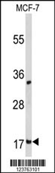
Western blot analysis of DSTN Antibody (Center) in MCF-7 cell line lysates (35 ug/lane). DSTN (arrow) was detected using the purified Pab.
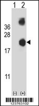
Western blot analysis of DSTN (arrow) using rabbit polyclonal DSTN Antibody (Center). 293 cell lysates (2 ug/lane) either nontransfected (Lane 1) or transiently transfected (Lane 2) with the DSTN gene.
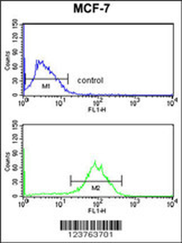
DSTN Antibody (Center) flow cytometry analysis of MCF-7 cells (bottom histogram) compared to a negative control cell (top histogram). FITC-conjugated goat-anti-rabbit secondary antibodies were used for the analysis.
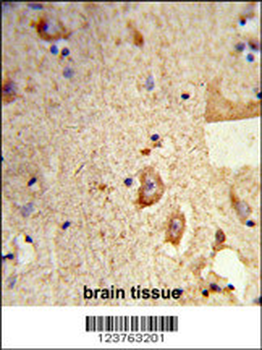
Formalin-fixed and paraffin-embedded human brain tissue reacted with DSTN Antibody (Center), which was peroxidase-conjugated to the secondary antibody, followed by DAB staining. This data demonstrates the use of this antibody for immunohistochemistry; clinical relevance has not been evaluated.
