You have no items in your shopping cart.
Cart summary
Item 1 of 1
Cytokeratin 19 antibody (FITC)
Catalog Number: orb154471
| Catalog Number | orb154471 |
|---|---|
| Category | Antibodies |
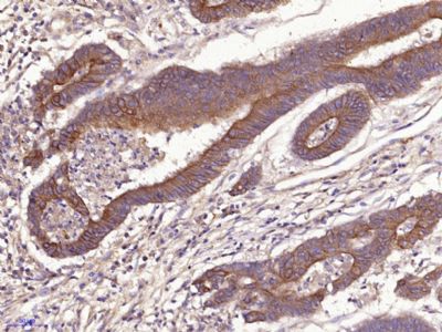
| Description | Mouse monoclonal antibody conjugated to FITC which recognizes Cytokeratin 19 that belongs to the intermediate filament family. It expressed in a defined zone of basal keratinocytes in the deep outer root sheath of hair follicles. It involved in the organization of myofibers. Cytokeratin 19 is a specific marker of moderate to severe dysplasia and carcinoma in situ in oral cavity squamous epithelium, and measurement of Cytokeratin 19 may be a useful marker in diagnosing hepatoma. |
| Clonality | Monoclonal |
| Clone Number | A53-B/A2 |
| Tested applications | FC |
| Reactivity | Human |
| Isotype | Mouse IgG2a |
| Immunogen | MCF-7 human breast adenocarcinoma cell line |
| Concentration | 1 mg/ml |
| Dilution range | Flow cytometry: Recommended dilution: 1-5 μg/ml. Intracellular staining. |
| Purity | Purified antibody is conjugated with fluorescein isothiocyanate (FITC) under optimum conditions and unconjugated antibody and free fluorochrome are removed by size-exclusion chromatography. |
| Conjugation | FITC |
| Target | Cytokeratin 19 |
| Entrez | 3880 |
| UniProt ID | P08727 |
| Storage | Store at 2-8°C. Protect from prolonged exposure to light. Do not freeze. |
| Buffer/Preservatives | Phosphate buffered saline (PBS), pH 7.4, 15 mM sodium azide |
| Alternative names | Anti-K19 antibody, anti-CK19 antibody, anti-CYK19 Read more... |
| Note | For research use only |
| Application notes | Flow cytometry: Recommended dilution: 1-5 μg/ml. Intracellular staining. |
| Expiration Date | 12 months from date of receipt. |
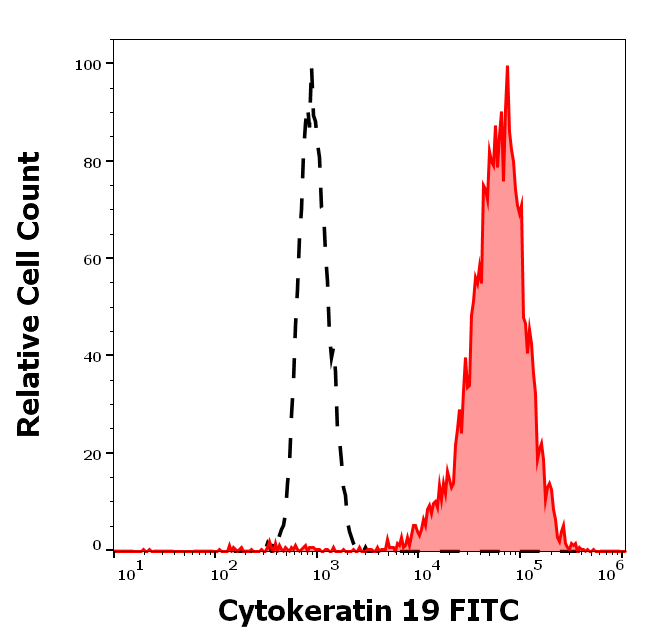
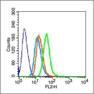
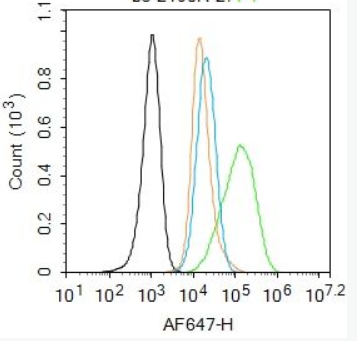
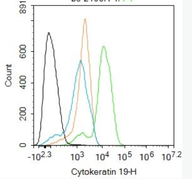
Separation of MCF-7 cells stained using anti-Cytokeratin 19 (A53-B/A2) FITC antibody (concentration in sample 3 µg/ml, red-filled) from MCF-7 cells stained using mouse IgG2b isotype control (MPC-11) FITC antibody (concentration in sample 3 µg/ml, same as anti-Cytokeratin 19 FITC antibody concentration, black-dashed) in flow cytometry analysis (surface staining).
- Item 1 of 11

Submit a review
Filter by Rating

- 5 stars

- 4 stars
- 3 stars
- 2 stars
- 1 stars