You have no items in your shopping cart.
Cart summary
Item 1 of 7
Item 1 of 7
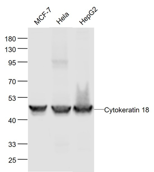
CYK18 Antibody
Catalog Number: orb1263271

| Catalog Number | orb1263271 |
|---|---|
| Category | Antibodies |
| Description | CYK18 Antibody |
| Target | KRT18 |
| Clonality | Polyclonal |
| Isotype | Rabbit Ig |
| Conjugation | Unconjugated |
| Reactivity | Human, Mouse |
| Form/Appearance | Liquid |
| Concentration | batch dependent |
| Buffer/Preservatives | Supplied in PBS with 0.09% (W/V) sodium azide. |
| Immunogen | This CYK18 antibody is generated from rabbits immunized with a KLH conjugated synthetic peptide between 401-430 amino acids from the C-terminal region of human CYK18. |
| UniProt ID | P05783 |
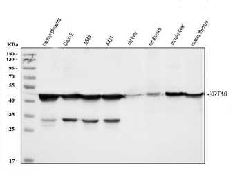
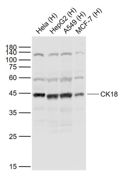
| MW | 48 kDa |
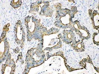
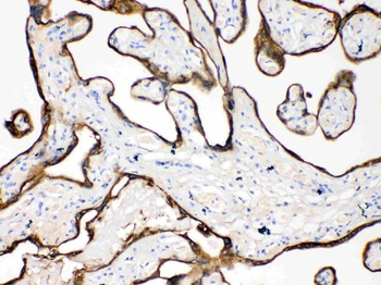
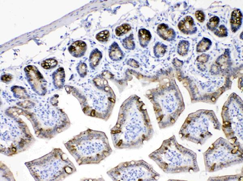
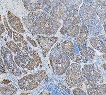
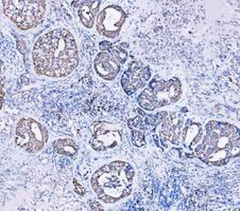
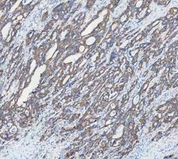
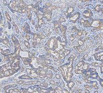
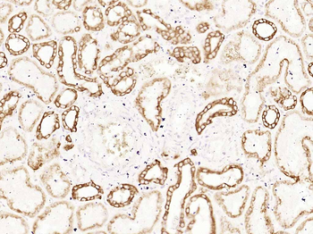
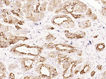
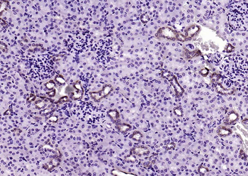
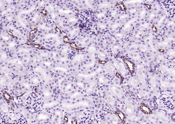
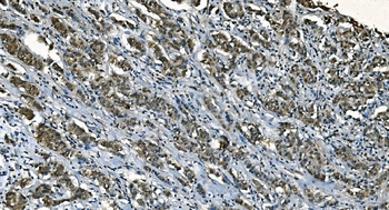
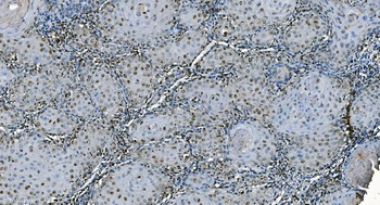
| Tested applications | FC, IF, IHC-P, WB |
| Application notes | For IF starting dilution is: 1:25For FACS starting dilution is: 1:25 |
| Antibody Type | Primary Antibody |
| Storage | Maintain refrigerated at 2-8°C for up to 2 weeks. For long term storage store at -20°C in small aliquots to prevent freeze-thaw cycles. |
| Alternative names | Keratin, type I cytoskeletal 18, Cell proliferatio Read more... |
| Note | For research use only |
| NCBI | P05783 |
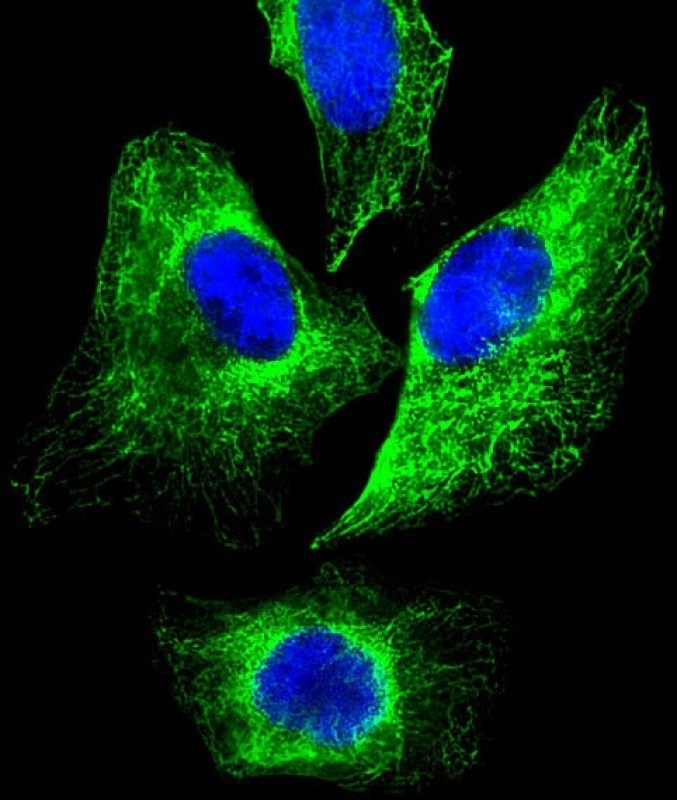
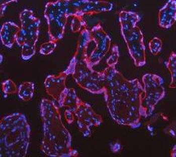
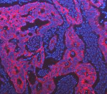
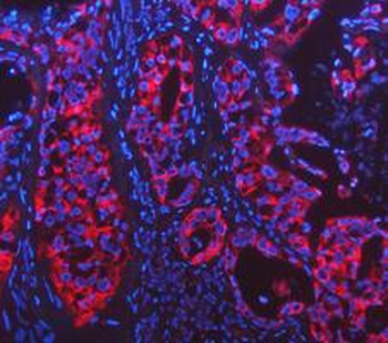
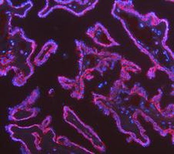
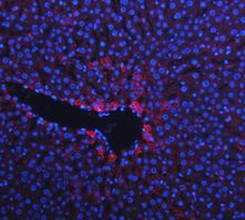
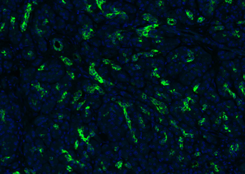
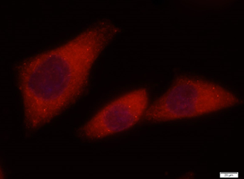
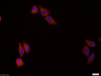
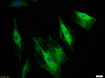
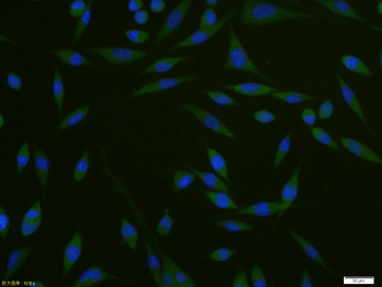
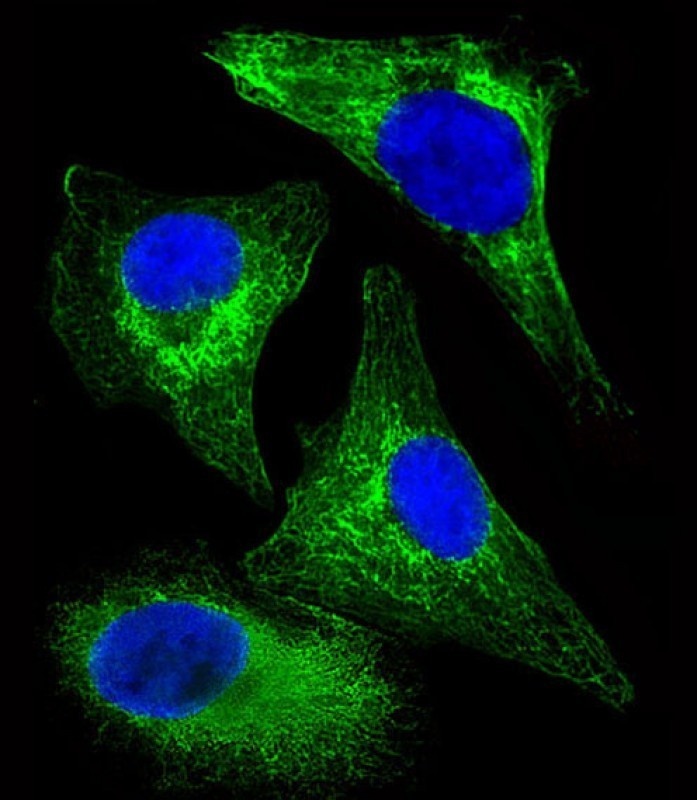
Immunofluorescent analysis of 4% paraformaldehyde-fixed, 0.1% Triton X-100 permeabilized HeLa (human cervical epithelial adenocarcinoma cell line) cells labeling Pdx1 with antibody at 1/25 dilution, followed by 488-conjugated goat anti-rabbit IgG secondary antibody at 1/200 dilution (green). Immunofluorescence image showing cytoskeleton staining on HeLa cell line. The nuclear counter stain is DAPI (blue).
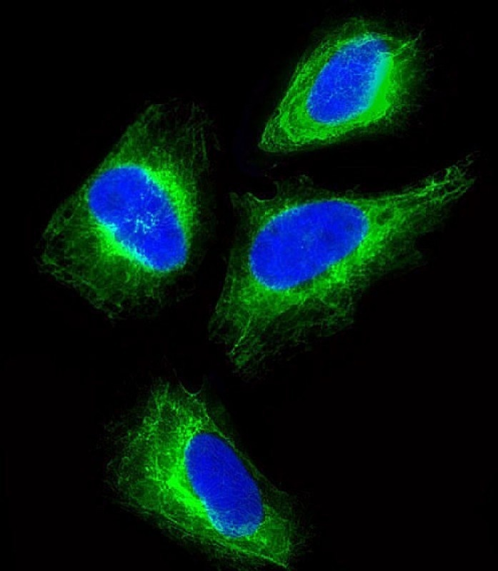
Immunofluorescent analysis of 4% paraformaldehyde-fixed, 0.1% Triton X-100 permeabilized HeLa (human cervical epithelial adenocarcinoma cell line) cells labeling Pdx1 with antibody at 1/25 dilution, followed by 488-conjugated goat anti-rabbit IgG secondary antibody at 1/200 dilution (green). Immunofluorescence image showing cytoskeleton staining on HeLa cell line. The nuclear counter stain is DAPI (blue).
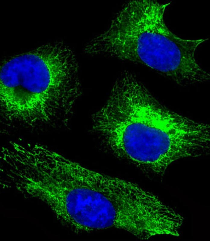
Immunofluorescent analysis of 4% paraformaldehyde-fixed, 0.1% Triton X-100 permeabilized HeLa (human cervical epithelial adenocarcinoma cell line) cells labeling Pdx1 with antibody at 1/25 dilution, followed by 488-conjugated goat anti-rabbit IgG secondary antibody at 1/200 dilution (green). Immunofluorescence image showing cytoskeleton staining on HeLa cell line. The nuclear counter stain is DAPI (blue).
Immunofluorescent analysis of 4% paraformaldehyde-fixed, 0.1% Triton X-100 permeabilized HeLa (human cervical epithelial adenocarcinoma cell line) cells labeling Pdx1 with antibody at 1/25 dilution, followed by 488-conjugated goat anti-rabbit IgG secondary antibody at 1/200 dilution (green). Immunofluorescence image showing cytoskeleton staining on HeLa cell line. The nuclear counter stain is DAPI (blue).
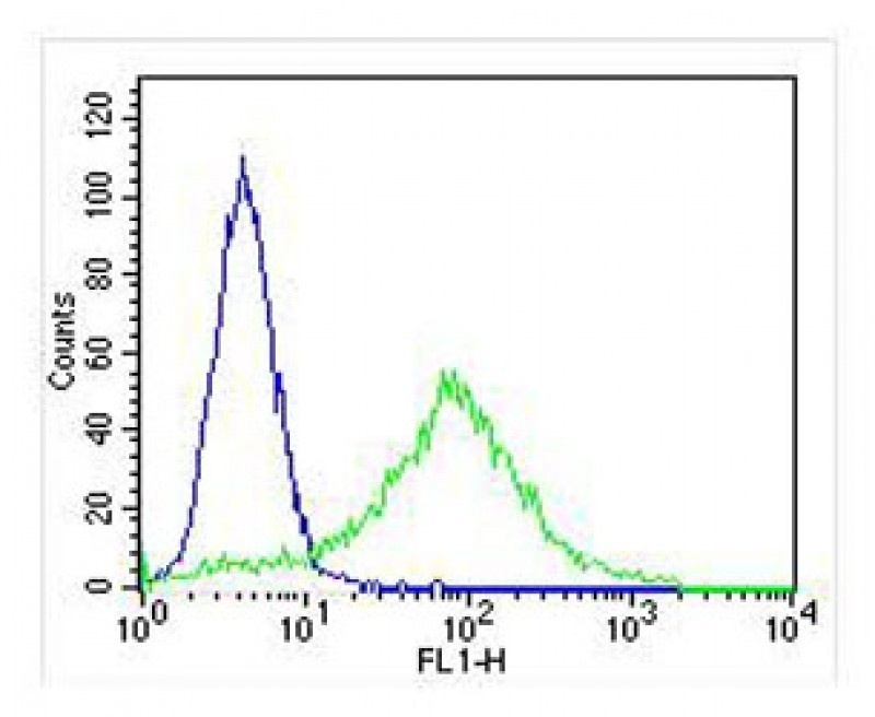
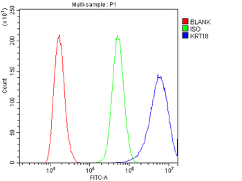
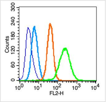
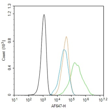
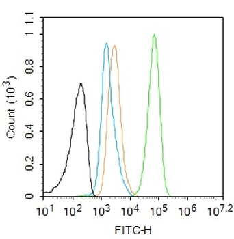
Overlay histogram showing Hela cells stained with Antibody (green line). The cells were fixed with 2% paraformaldehyde (10 min) and then permeabilized with 90% methanol for 10 min. The cells were then icubated in 2% bovine serum albumin to block non-specific protein-protein interactions followed by the antibody (1:25 dilution) for 60 min at 37°C. The secondary antibody used was Goat-Anti-Rabbit IgG, Conjugated Highly Cross-Adsorbed at 1/400 dilution for 40 min at 37°C. Isotype control antibody (blue line) was rabbit IgG (1ug/1x10^6 cells) used under the same conditions. Acquisition of > 10000 events was performed.
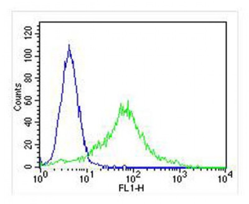
Overlay histogram showing Hela cells stained with Antibody (green line). The cells were fixed with 2% paraformaldehyde (10 min) and then permeabilized with 90% methanol for 10 min. The cells were then icubated in 2% bovine serum albumin to block non-specific protein-protein interactions followed by the antibody (1:25 dilution) for 60 min at 37°C. The secondary antibody used was Goat-Anti-Rabbit IgG, Conjugated Highly Cross-Adsorbed at 1/400 dilution for 40 min at 37°C. Isotype control antibody (blue line) was rabbit IgG (1ug/1x10^6 cells) used under the same conditions. Acquisition of > 10000 events was performed.
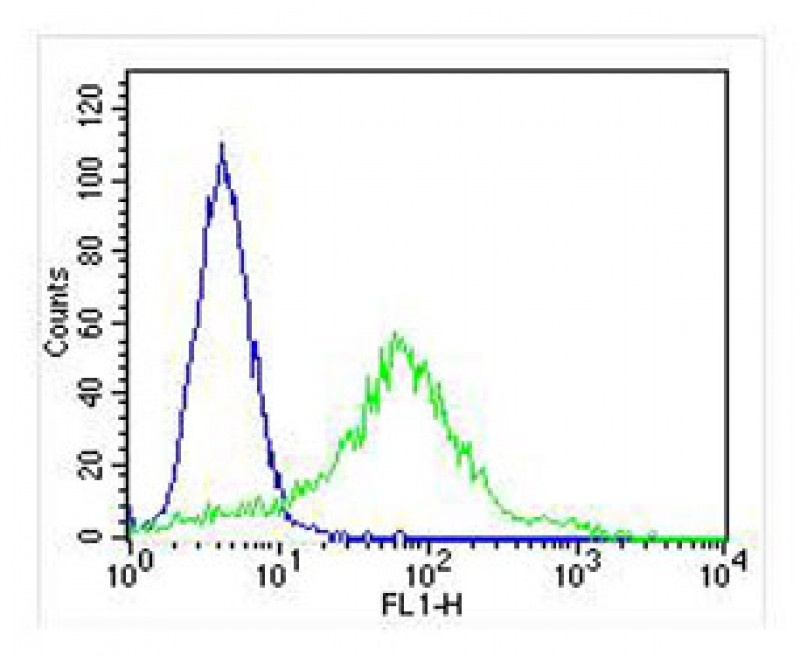
Overlay histogram showing Hela cells stained with Antibody (green line). The cells were fixed with 2% paraformaldehyde (10 min) and then permeabilized with 90% methanol for 10 min. The cells were then icubated in 2% bovine serum albumin to block non-specific protein-protein interactions followed by the antibody (1:25 dilution) for 60 min at 37°C. The secondary antibody used was Goat-Anti-Rabbit IgG, Conjugated Highly Cross-Adsorbed at 1/400 dilution for 40 min at 37°C. Isotype control antibody (blue line) was rabbit IgG (1ug/1x10^6 cells) used under the same conditions. Acquisition of > 10000 events was performed.
- Item 1 of 21
Anti-Cytokeratin 18/KRT18 Antibody [orb389489]
FC, ICC, IF, IHC, IHC-Fr, WB
Human, Mouse, Rat
Rabbit
Polyclonal
Unconjugated
10 μg, 100 μg - Item 1 of 12
CK18 Mouse Monoclonal Antibody [orb499665]
ICC, IF, IHC-Fr, IHC-P, WB
Mouse, Rat
Human, Mouse, Rat
Mouse
Monoclonal
Unconjugated
200 μg, 200 μl, 100 μl, 50 μl - Item 1 of 8
CK18 Rabbit Polyclonal Antibody [orb5871]
FC, ICC, IF, IHC-Fr, IHC-P, WB
Canine, Gallus, Mouse, Rabbit
Human, Rat
Rabbit
Polyclonal
Unconjugated
100 μl, 200 μl, 50 μl - Item 1 of 10
CK18 Recombinant Rabbit Monoclonal Antibody [orb500277]
ICC, IF, IHC-Fr, IHC-P, WB
Mouse, Rat
Human, Mouse, Rat
Rabbit
Recombinant
Unconjugated
50 μl, 100 μl - Item 1 of 9
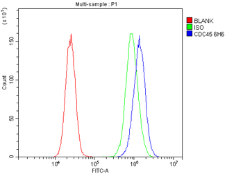
Anti-CDC45L Antibody (monoclonal, 6H6) [orb738407]
FC, IHC, WB
Human, Mouse, Rat
Mouse
Monoclonal
Unconjugated
100 μg, 10 μg