You have no items in your shopping cart.
Cart summary
Item 1 of 5
Item 1 of 5
CTSA Antibody
Catalog Number: orb1270799

| Catalog Number | orb1270799 |
|---|---|
| Category | Antibodies |
| Description | CTSA Antibody |
| Target | CTSA |
| Clonality | Polyclonal |
| Isotype | Rabbit Ig |
| Conjugation | Unconjugated |
| Reactivity | Human, Mouse |
| Form/Appearance | Liquid |
| Concentration | batch dependent |
| Buffer/Preservatives | Supplied in PBS with 0.09% (W/V) sodium azide. |
| Immunogen | This CTSA antibody is generated from rabbits immunized with a KLH conjugated synthetic peptide between 18-45 amino acids from the N-terminal region of human CTSA. |
| UniProt ID | P10619 |
| MW | 54 kDa |
| Tested applications | IHC-P, WB |
| Application notes | For IHC-P starting dilution is: 1:25For WB starting dilution is: 1:1000 |
| Antibody Type | Primary Antibody |
| Storage | Maintain refrigerated at 2-8°C for up to 2 weeks. For long term storage store at -20°C in small aliquots to prevent freeze-thaw cycles. |
| Alternative names | Lysosomal protective protein, Carboxypeptidase C, Read more... |
| Note | For research use only |
| NCBI | P10619 |
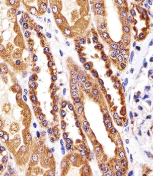
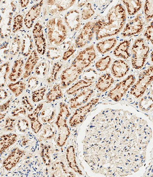
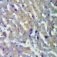
Immunohistochemical analysis of paraffin-embedded H. kidney section using CTSA Antibody (N-term). Antibody was diluted at 1:25 dilution. A undiluted biotinylated goat polyvalent antibody was used as the secondary, followed by DAB staining.

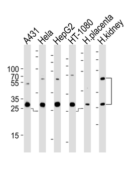
Western blot analysis of lysates from A431, Hela, HepG2, HT-1080 cell line and human placenta, kidney tissue lysate (from left to right), using CTSA Antibody at 1:1000 at each lane.

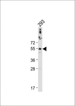
Western blot analysis in HepG2 cell line lysates (35 ug/lane).

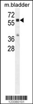
Western blot analysis in mouse bladder tissue lysates (35 ug/lane).
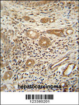
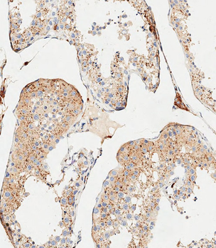
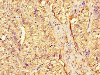
CTSA antibody immunohistochemistry analysis in formalin fixed and paraffin embedded human hepatocarcinoma followed by peroxidase conjugation of the secondary antibody and DAB staining.
- Item 1 of 6
- Item 1 of 1
- Item 1 of 1
- Item 1 of 3
Anti-Cathepsin A 32k Antibody [orb214425]
IF, IH, WB
Human, Mouse, Rat
Rabbit
Polyclonal
Unconjugated
30 μl, 100 μl, 200 μl - Item 1 of 3