You have no items in your shopping cart.
Cart summary
Item 1 of 5
Item 1 of 5
CSE1L Antibody
Catalog Number: orb1265562
| Catalog Number | orb1265562 |
|---|---|
| Category | Antibodies |
| Description | CSE1L Antibody |
| Species/Host | Rabbit |
| Clonality | Polyclonal |
| Tested applications | IF, IHC-P, WB |
| Predicted Reactivity | Bovine, Zebrafish |
| Reactivity | Human, Mouse |
| Isotype | Rabbit Ig |
| Immunogen | This Cellular Apoptosis Susceptibility antibody is generated from rabbits immunized with a KLH conjugated synthetic peptide between 55-84 amino acids from the N-terminal region of human Cellular Apoptosis Susceptibility. |
| Concentration | batch dependent |
| Dilution range | For IHC-P starting dilution is: 1:100For IF starting dilution is: 1:100For WB starting dilution is: 1:1000 |
| Form/Appearance | Liquid |
| Conjugation | Unconjugated |
| MW | 110 kDa |
| Target | CSE1L |
| UniProt ID | P55060 |
| NCBI | P55060 |
| Storage | Store at 4°C for three months and -20°C, stable for up to one year. As with all antibodies care should be taken to avoid repeated freeze thaw cycles. Antibodies should not be exposed to prolonged high temperatures. |
| Buffer/Preservatives | Supplied in PBS with 0.09% (W/V) sodium azide. |
| Alternative names | Exportin-2, Exp2, Cellular apoptosis susceptibilit Read more... |
| Note | For research use only |
| Application notes | For IHC-P starting dilution is: 1:100For IF starting dilution is: 1:100For WB starting dilution is: 1:1000 |
| Expiration Date | 12 months from date of receipt. |

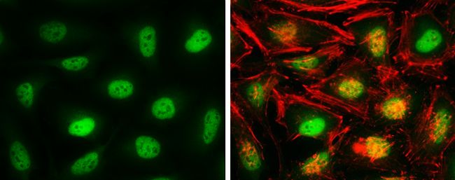
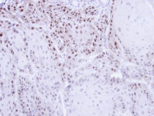
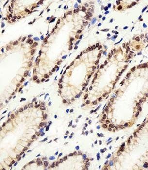
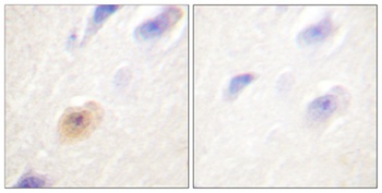
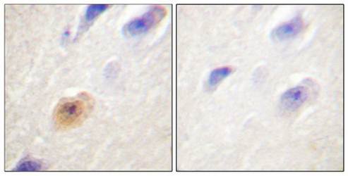
Immunohistochemical analysis of paraffin-embedded H. stomach section using CSE1L Antibody. Antibody was diluted at 1:100 dilution. A peroxidase-conjugated goat anti-rabbit IgG at 1:400 dilution was used as the secondary antibody, followed by DAB staining.
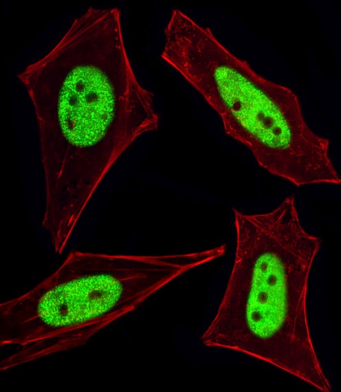
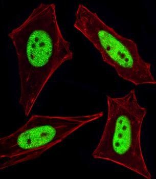
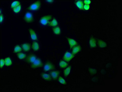
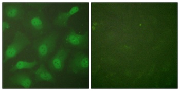
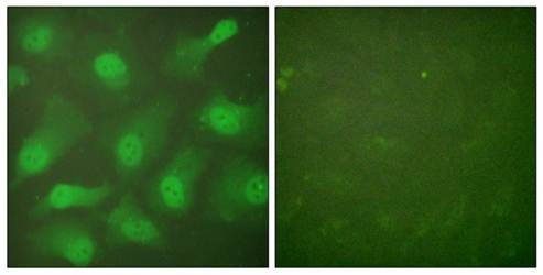
Fluorescent image of HeLa cells stained with Cellular Apoptosis Susceptibility Antibody. An Alexa Fluor 488-conjugated goat anti-rabbit lgG at 1:400 dilution was used as the secondary antibody (green). Cytoplasmic actin was counterstained with Alexa Fluor 555 conjugated with Phalloidin (red).
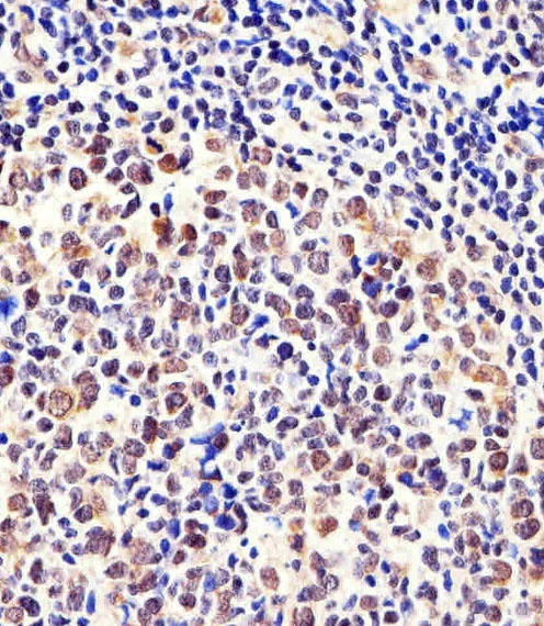
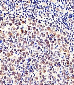
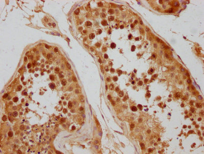
Immunohistochemical analysis of paraffin-embedded H. tonsil section using CSE1L Antibody. Antibody was diluted at 1:100 dilution. A peroxidase-conjugated goat anti-rabbit IgG at 1:400 dilution was used as the secondary antibody, followed by DAB staining.
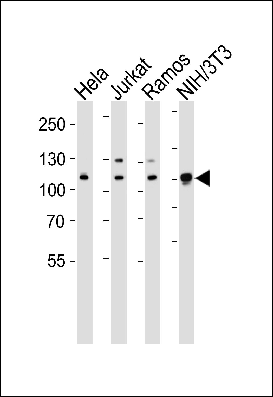
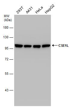
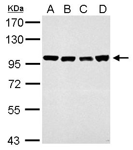
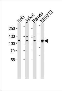
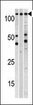
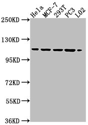
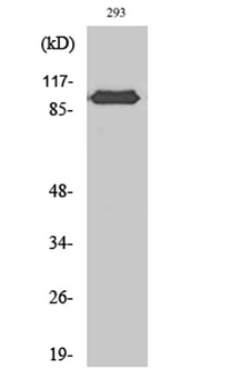
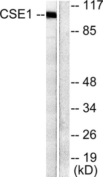
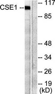
Western blot analysis of lysates from Hela, Jurkat, Ramos, mouse NIH/3T3 cell line (from left to right), using CSE1L Antibody at 1:1000 at each lane.

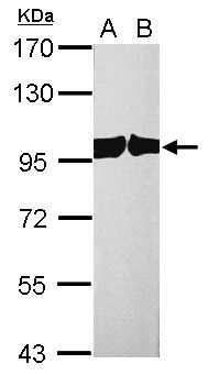
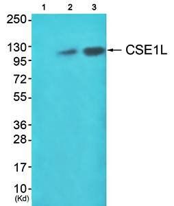
Western blot analysis of anti-CSE1L Pab in, from left to right, A375, CEM, and mouse heart cell line lysates (35 ug/lane).
- Item 1 of 5
chromosome segregation 1 like Antibody [orb556066]
ICC, IHC-P, IP, WB
Human, Mouse, Rat
Rabbit
Polyclonal
Unconjugated
100 μl - Item 1 of 5
Cellular Apoptosis Susceptibility antibody [orb33490]
IF, IHC-P, WB
Zebrafish
Human, Mouse
Rabbit
Polyclonal
Unconjugated
80 μl - Item 1 of 3
- Item 1 of 4
- Item 1 of 4
CSE1L antibody [orb125620]
IF, IHC, IP, WB
Human
Rabbit
Polyclonal
Unconjugated
200 μg, 100 μg, 50 μg, 25 μg
Submit a review
Filter by Rating

- 5 stars

- 4 stars
- 3 stars
- 2 stars
- 1 stars