You have no items in your shopping cart.
Cart summary
Item 1 of 17
Item 1 of 17
COX5B Antibody
Catalog Number: orb1173497
| Catalog Number | orb1173497 |
|---|---|
| Category | Antibodies |
| Description | COX5B Antibody |
| Species/Host | Rabbit |
| Clonality | Polyclonal |
| Tested applications | ELISA, FC, ICC, IF, IHC, WB |
| Reactivity | Human, Mouse, Rat |
| Isotype | Rabbit IgG |
| Immunogen | E.coli-derived human COX5B recombinant protein (Position: M1-H129). |
| Concentration | Adding 0.2 ml of distilled water will yield a concentration of 500 μg/ml. |
| Dilution range | Western blot, 0.1-0.25 μg/ml, Human, Mouse, Rat Immunohistochemistry(Paraffin-embedded Section), 2-5 μg/ml, Human, Mouse, Rat Immunocytochemistry/Immunofluorescence, 5 μg/ml, Human Immunofluorescence, 5 μg/ml, Human Flow Cytometry, 1-3 μg/1x10^6 cells, Human Direct ELISA, 0.1-0.5 μg/ml, Human |
| Form/Appearance | Lyophilized |
| Conjugation | Unconjugated |
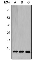
| MW | 15 kDa |
| UniProt ID | P10606 |
| Storage | At -20°C for one year from date of receipt. After reconstitution, at 4°C for one month. It can also be aliquotted and stored frozen at -20°C for six months. Avoid repeated freezing and thawing. |
| Note | For research use only |
| Application notes | Tested Species: In-house tested species with positive results. Other applications have not been tested. Optimal dilutions should be determined by end users. Adding 0.2 ml of distilled water will yield a concentration of 500 μg/ml. |
| Expiration Date | 12 months from date of receipt. |
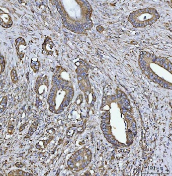
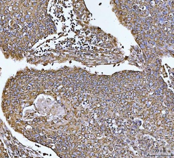
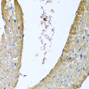
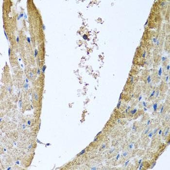
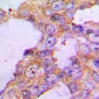
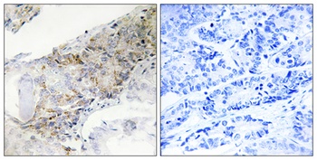
IHC analysis of COX5B using anti-COX5B antibody. COX5B was detected in a paraffin-embedded section of human breast cancer tissue.
IHC analysis of COX5B using anti-COX5B antibody. COX5B was detected in a paraffin-embedded section of human endometrial carcinoma tissue.
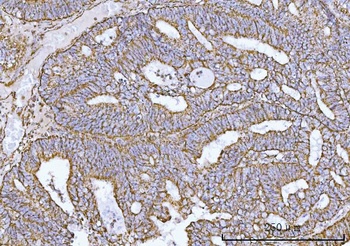
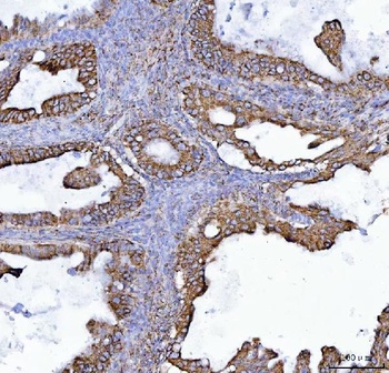
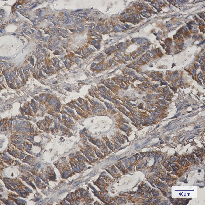
IHC analysis of COX5B using anti-COX5B antibody. COX5B was detected in a paraffin-embedded section of human colorectal adenocarcinoma tissue.
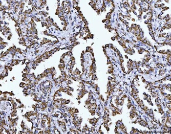
IHC analysis of COX5B using anti-COX5B antibody. COX5B was detected in a paraffin-embedded section of human lung cancer tissue.
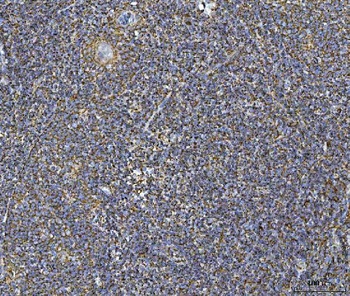
IHC analysis of COX5B using anti-COX5B antibody. COX5B was detected in a paraffin-embedded section of human tonsil tissue.
IHC analysis of COX5B using anti-COX5B antibody. COX5B was detected in a paraffin-embedded section of human esophageal squamous carcinoma tissue.
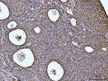
IHC analysis of COX5B using anti-COX5B antibody. COX5B was detected in a paraffin-embedded section of human ovarian cancer tissue.
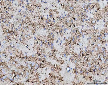
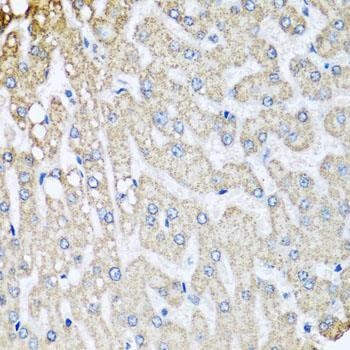
IHC analysis of COX5B using anti-COX5B antibody. COX5B was detected in a paraffin-embedded section of mouse brain tissue.
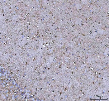
IHC analysis of COX5B using anti-COX5B antibody. COX5B was detected in a paraffin-embedded section of mouse brain tissue.
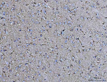
IHC analysis of COX5B using anti-COX5B antibody. COX5B was detected in a paraffin-embedded section of rat brain tissue.
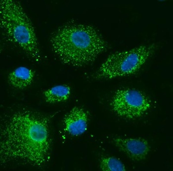
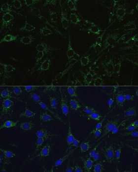
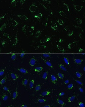
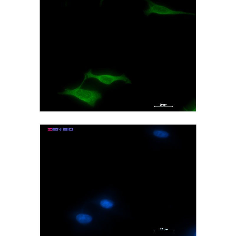
IF analysis of COX5B using anti-COX5B antibody. COX5B was detected in an immunocytochemical section of A549 cells.
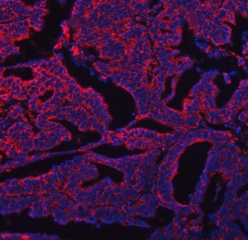
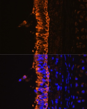
IF analysis of COX5B using anti-COX5B antibody. COX5B was detected in a paraffin-embedded section of human ovary cancer tissue.
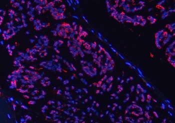
IF analysis of COX5B using anti-COX5B antibody. COX5B was detected in a paraffin-embedded section of human breast cancer tissue.
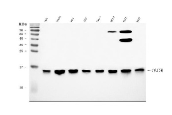
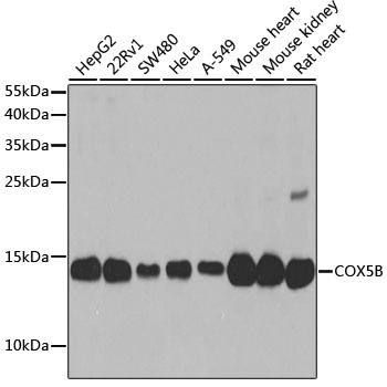
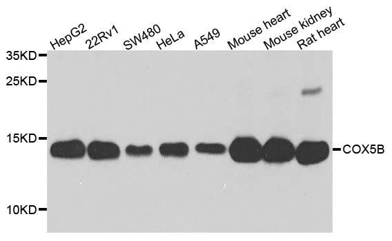
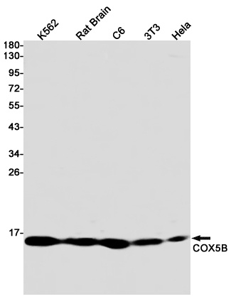
Western blot analysis of COX5B using anti-COX5B antibody.
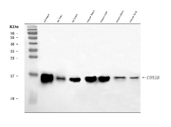
Western blot analysis of COX5B using anti-COX5B antibody.
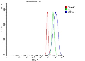
Flow Cytometry analysis of HeLa cells using anti-COX5B antibody(Blue line).Isotype control antibody(Green line) was rabbit IgG.Unlabelled sample(Red line) was also used as a control.
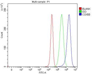
Flow Cytometry analysis of 293T cells using anti-COX5B antibody(Blue line).Isotype control antibody(Green line) was rabbit IgG.Unlabelled sample(Red line) was also used as a control.
- Item 1 of 6
- Item 1 of 3
COX5B antibody [orb135301]
ICC, IF, IHC, IP, WB
Human, Mouse, Rat
Polyclonal
Unconjugated
50 μl, 100 μl, 200 μl - Item 1 of 3
COX5B Antibody [orb1565059]
ICC, IHC-Fr, IHC-P, IP, WB
Human, Mouse, Rat
Rabbit
Monoclonal
Unconjugated
50 μl, 20 μl, 100 μl - Item 1 of 2
COX5B antibody [orb235111]
IH, WB
Human, Mouse, Porcine, Rat
Rabbit
Polyclonal
Unconjugated
200 μl, 100 μl, 30 μl - Item 1 of 2

Submit a review
Filter by Rating

- 5 stars

- 4 stars
- 3 stars
- 2 stars
- 1 stars