You have no items in your shopping cart.
Cart summary
Item 1 of 3
Item 1 of 3
CLCN4 Antibody
Catalog Number: orb1263869
| Catalog Number | orb1263869 |
|---|---|
| Category | Antibodies |
| Description | CLCN4 Antibody |
| Species/Host | Rabbit |
| Clonality | Polyclonal |
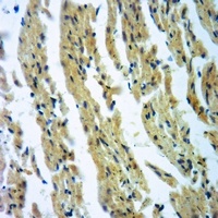
| Tested applications | IF, IHC-P, WB |
| Predicted Reactivity | Mouse, Rat |
| Reactivity | Human |
| Isotype | Rabbit Ig |
| Immunogen | This CLC4 antibody is generated from rabbits immunized with a KLH conjugated synthetic peptide between 663-689 amino acids from the C-terminal region of human CLC4. |
| Concentration | batch dependent |
| Dilution range | For WB starting dilution is: 1:1000For IF starting dilution is: 1:10~50For IHC-P starting dilution is: 1:10~50 |
| Form/Appearance | Liquid |
| Conjugation | Unconjugated |
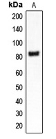
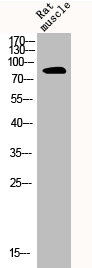
| MW | 85 kDa |
| Target | CLCN4 |
| UniProt ID | P51793 |
| NCBI | P51793 |
| Storage | Store at 4°C for three months and -20°C, stable for up to one year. As with all antibodies care should be taken to avoid repeated freeze thaw cycles. Antibodies should not be exposed to prolonged high temperatures. |
| Buffer/Preservatives | Supplied in PBS with 0.09% (W/V) sodium azide. |
| Alternative names | H(+)/Cl(-) exchange transporter 4, Chloride channe Read more... |
| Note | For research use only |
| Application notes | For WB starting dilution is: 1:1000For IF starting dilution is: 1:10~50For IHC-P starting dilution is: 1:10~50 |
| Expiration Date | 12 months from date of receipt. |
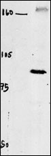
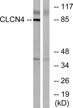
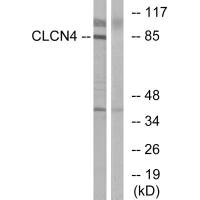
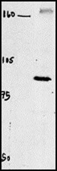
Western blot of chicken brain tissue incubated with CLC4 Antibody.

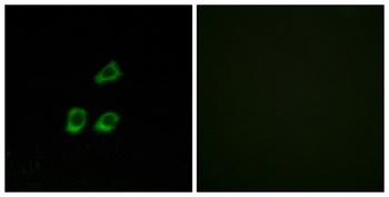
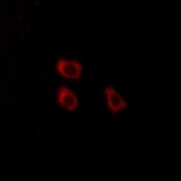
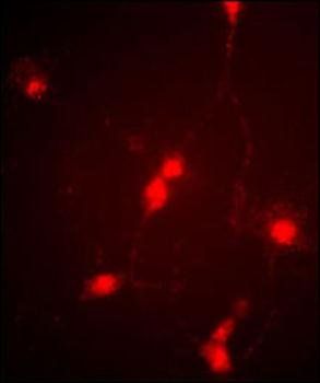
Immunofluorescence image of cultured chick retinal amacrine (neuronal) cells labeled with CLC4 Antibody.
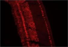
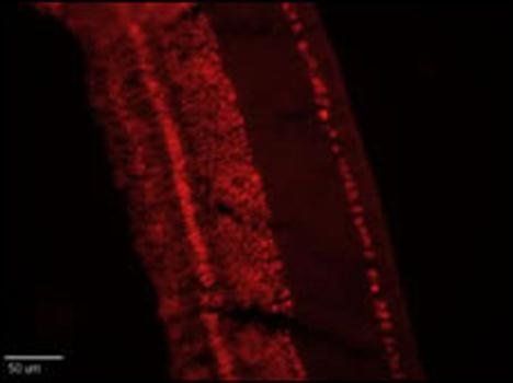
Retinae were collected from adult White Leghorn chicken and fixed in the eyecup for two hours in 4% Paraformaldehyde (in PBS). Retinae were then removed from the eyecups and incubated in 30% sucrose (in PBS) overnight. Retinal tissue was embedded and frozen in OCT compound and cut into ~15um sections on a cryotome. Sections were blocked in 5% normal goat serum (in 1%BSA/.1%saponin PBS) for one hour and then incubated at RT with 1:250 (1%BSA/.1% saponin PBS) ClC4 antibody for 1 hour. Sections were then washed in PBS (3X10minutes) and then treated with secondary antibody (1:500 Cy3) for one hour. After another PBS wash series, sections were coverslipped and antibody labeling was visualized at 20X with a Leica upright microscope using a TRITC filter set and Xenon lamp illumination.
- Item 1 of 4
- Item 1 of 3
CLCN4 antibody [orb213747]
IF, IH, WB
Human, Mouse, Primate, Rat, Zebrafish
Rabbit
Polyclonal
Unconjugated
200 μl, 100 μl, 30 μl - Item 1 of 2
- Item 1 of 2
- Item 1 of 3
Submit a review
Filter by Rating

- 5 stars

- 4 stars
- 3 stars
- 2 stars
- 1 stars