You have no items in your shopping cart.
Cart summary
Item 1 of 5
Item 1 of 5
CDH4 Antibody
Catalog Number: orb1268616

| Catalog Number | orb1268616 |
|---|---|
| Category | Antibodies |
| Description | CDH4 Antibody |
| Species/Host | Rabbit |
| Clonality | Polyclonal |
| Tested applications | FC, IF, IHC-P, WB |
| Predicted Reactivity | Mouse, Rat |
| Reactivity | Human |
| Isotype | Rabbit Ig |
| Immunogen | This CDH4 antibody is generated from rabbits immunized with a KLH conjugated synthetic peptide between 175-203 amino acids from the N-terminal region of human CDH4. |
| Antibody Type | Primary Antibody |
| Concentration | batch dependent |
| Form/Appearance | Liquid |
| Conjugation | Unconjugated |
| MW | 100 kDa |
| Target | CDH4 |
| UniProt ID | P55283 |
| NCBI | P55283 |
| Storage | Maintain refrigerated at 2-8°C for up to 2 weeks. For long term storage store at -20°C in small aliquots to prevent freeze-thaw cycles. |
| Buffer/Preservatives | Supplied in PBS with 0.09% (W/V) sodium azide. |
| Alternative names | Cadherin-4, Retinal cadherin, R-CAD, R-cadherin, C Read more... |
| Note | For research use only |
| Application notes | For WB starting dilution is: 1:2000For IHC-P starting dilution is: 1:10~50For IF starting dilution is: 1:10~50For FACS starting dilution is: 1:10~50 |
| Expiration Date | 12 months from date of receipt. |
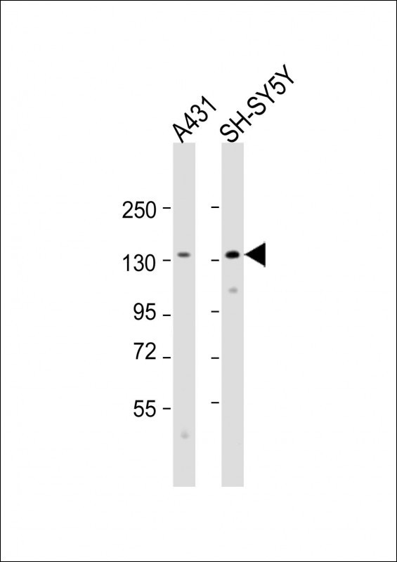
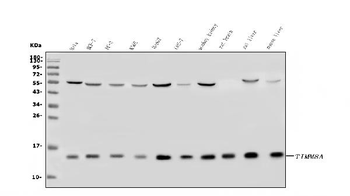
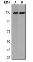
Western Blot at 1:2000 dilution Lane 1: A431 whole cell lysate Lane 2: SH-SY5Y whole cell lysate Lysates/proteins at 20 ug per lane.
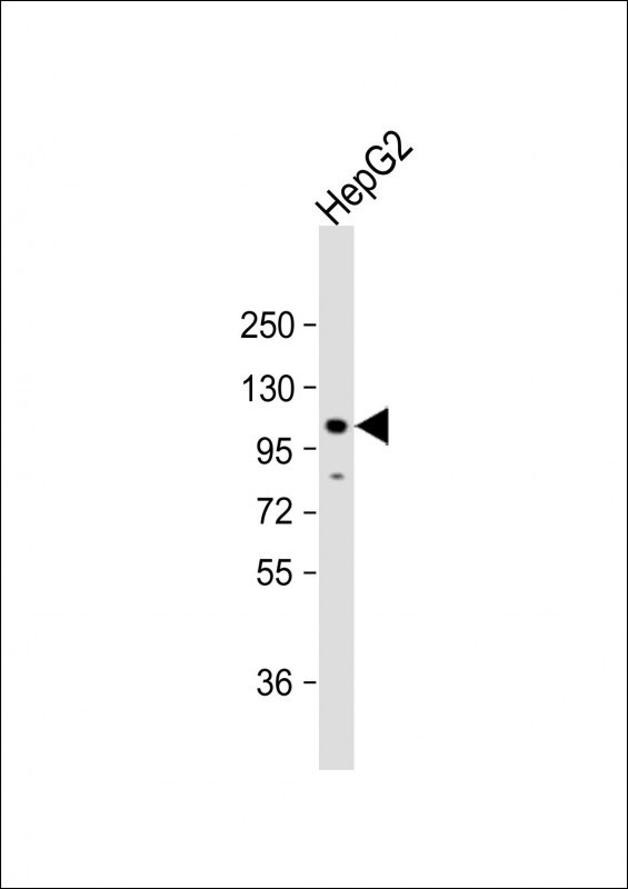
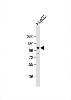
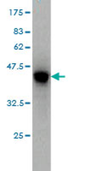
Western Blot at 1:2000 dilution + HepG2 whole cell lysate Lysates/proteins at 20 ug per lane.
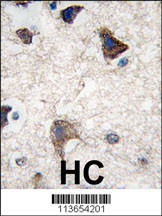
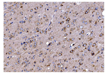
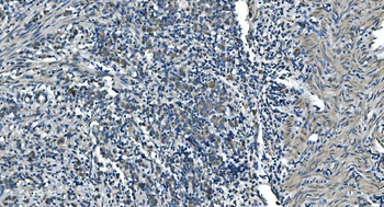
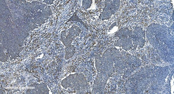
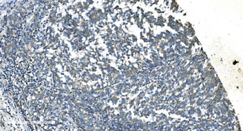
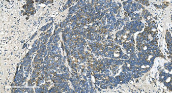
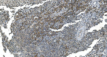
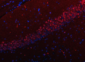
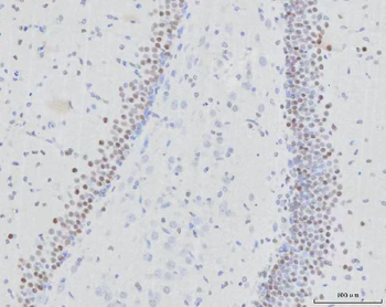
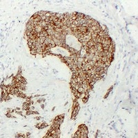
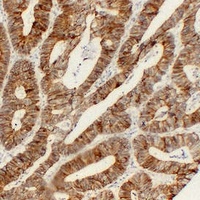
Formalin-fixed and paraffin-embedded human brain tissue reacted with CDH4 antibody, which was peroxidase-conjugated to the secondary antibody, followed by DAB staining.
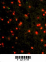
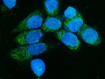
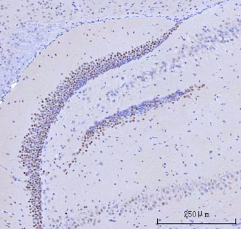
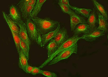
Immunofluorescence analysis of CDH4 Antibody Antibody with paraffin-embedded human brain tissue. 0.025 mg/ml primary antibody was followed by FITC-conjugated goat anti-rabbit lgG (whole molecule). FITC emits green fluorescence.Red counterstaining is PI.
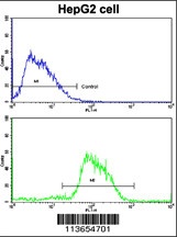
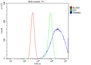
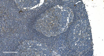
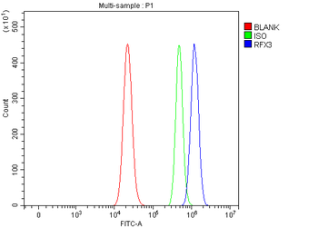
Flow cytometric analysis of HepG2 cells using CDH4 Antibody (N-term) (bottom histogram) compared to a negative control cell (top histogram). FITC-conjugated goat-anti-rabbit secondary antibodies were used for the analysis.
- Item 1 of 11
Anti-TIMM8A/DDP Antibody [orb670328]
ELISA, ICC, IF, IHC, WB
Human, Monkey, Mouse, Rat
Rabbit
Polyclonal
Unconjugated
10 μg, 100 μg - Item 1 of 6
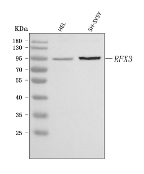
Anti-RFX3 Antibody [orb1676415]
ELISA, FC, ICC, IF, IHC, WB
Human, Mouse, Rat
Rabbit
Polyclonal
Unconjugated
10 μg, 100 μg - Item 1 of 5
CDH4 Antibody (N-term) [orb1936212]
FC, IF, IHC-P, WB
Mouse, Rat
Human
Rabbit
Polyclonal
Unconjugated
100 μl, 50 μl - Item 1 of 3
- Item 1 of 2
CDH4 monoclonal antibody (M01), clone 2E2 [orb2294670]
ELISA, WB
Human
Mouse
Monoclonal
Unconjugated
100 μg