You have no items in your shopping cart.
Cart summary
Item 1 of 3
Item 1 of 3
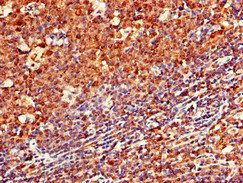
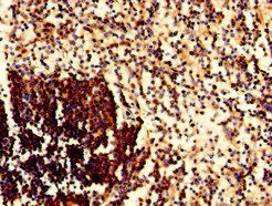
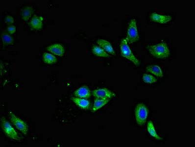
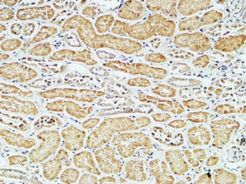
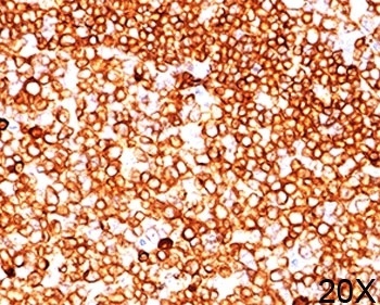
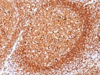
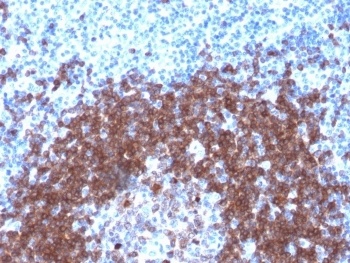
CD79a antibody
Catalog Number: orb154414
| Catalog Number | orb154414 |
|---|---|
| Category | Antibodies |
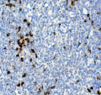
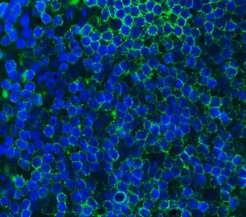
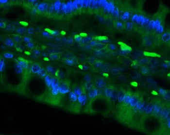
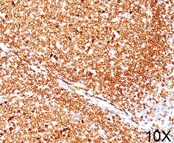
| Description | Mouse monoclonal antibody to CD79a which expressed in B-cells. Defects in CD79A are the cause of agammaglobulinemia type 3 (AGM3). The cytoplasmic tails of CD79A contains an ITAM (immuno-receptor tyrosine-based activation) motif, which acts to initiate the BCR signaling reactions by binding to and activating tyrosine |
| Clonality | Monoclonal |
| Clone Number | HM47 |
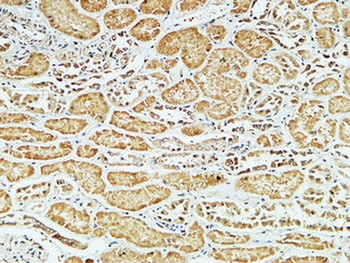
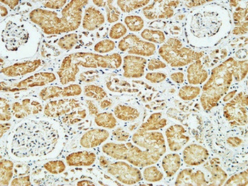
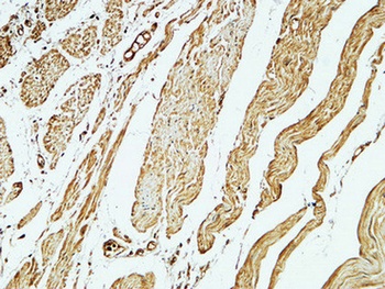
| Tested applications | FC, IHC-P, IP, WB |
| Reactivity | Bovine, Canine, Equine, Gallus, Guinea pig, Human, Mouse, Porcine, Primate, Rabbit, Rat |
| Isotype | Mouse IgG1 kappa |
| Immunogen | Synthetic peptide corresponding to C terminal amino acids 208-222 of human CD79a |
| Concentration | 1 mg/ml |
| Dilution range | Flow cytometry: Recommended dilution: 1-4 μg/ml, intracellular staining.Western blotting: Recommended dilution: 1-2 μg/ml. |
| Purity | Purified by protein-A affinity chromatography. |
| Conjugation | Unconjugated |
| Target | CD79a |
| Entrez | 973 |
| UniProt ID | P11912 |
| Storage | Store at 2-8°C. Do not freeze. |
| Buffer/Preservatives | Phosphate buffered saline (PBS), pH 7.4, 15 mM sodium azide |
| Alternative names | Anti-BCR alpha antibody, anti-Ig-alpha antibody, a Read more... |
| Note | For research use only |
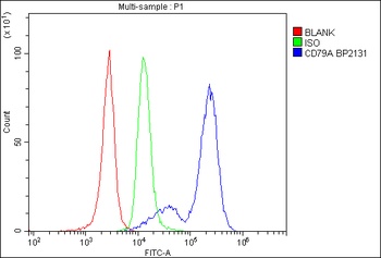
| Application notes | Flow cytometry: Recommended dilution: 1-4 μg/ml. Intracellular staining. |
| Expiration Date | 12 months from date of receipt. |
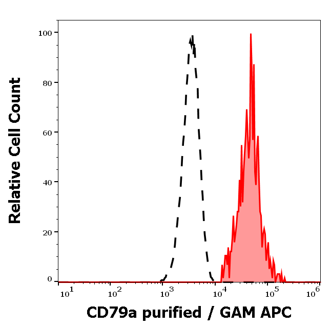
Separation of human CD79a positive lymphocytes (red-filled) from neutrophil granulocytes (black-dashed) in flow cytometry analysis (intracellular staining) of human peripheral whole blood stained using anti-human CD79a (HM47) purified antibody (concentration in sample 4 μg/ml, GAM APC).
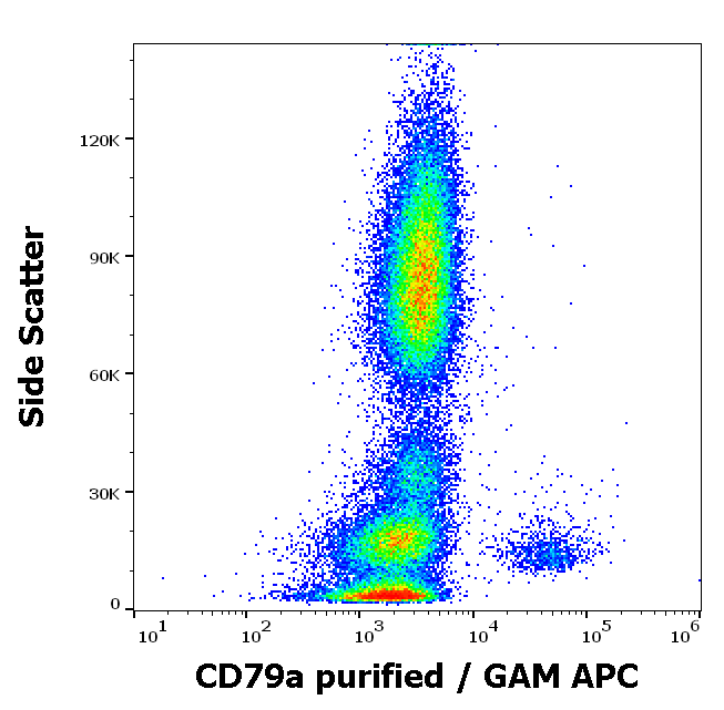
Flow cytometry intracellular staining pattern of human peripheral whole blood stained using anti-human CD79a (HM47) purified antibody (concentration in sample 4 μg/ml, GAM APC).

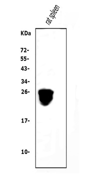
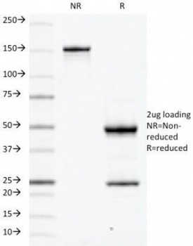
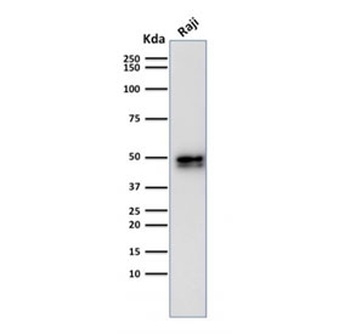
Western blotting analysis of human CD79a using mouse monoclonal antibody HM47 on lysates of Raji and Jurkat (negative control) cell line under reducing and non-reducing conditions. Nitrocellulose membrane was probed with 2 µg/ml of mouse anti-H-Ras monoclonal antibody followed by IRDye800-conjugated anti-mouse secondary antibody. CD79a was detected around 43 kDa.
- Item 1 of 6
Cd79a Antibody [orb654317]
ELISA, FC, IF, IHC, WB
Mouse, Rat
Rabbit
Polyclonal
Unconjugated
10 μg, 100 μg - Item 1 of 3
- Item 1 of 4
- Item 1 of 4
- Item 1 of 4
CD79A Antibody [orb1274259]
IHC-P, WB
Bovine, Human, Monkey, Mouse, Porcine, Rat
Rabbit
Recombinant
Unconjugated
100 μg
Submit a review
Filter by Rating

- 5 stars

- 4 stars
- 3 stars
- 2 stars
- 1 stars