You have no items in your shopping cart.
Cart summary
Item 1 of 3
Item 1 of 3
CD6 antibody
Catalog Number: orb44614
| Catalog Number | orb44614 |
|---|---|
| Category | Antibodies |
| Description | Mouse Monoclonal to CD6. |
| Clonality | Monoclonal |
| Clone Number | MEM-98 |
| Tested applications | FC, IHC-P, IP, WB |
| Reactivity | Human |
| Isotype | Mouse IgG1 |
| Immunogen | Human CD6 antigen purified by immunoaffinity chromatography from HBP-ALL cells followed by preparative SDS-PAGE of non-boiled non-reduced sample (excised piece of gel corresponding to the 100 kDa zone). |
| Concentration | 1 mg/ml |
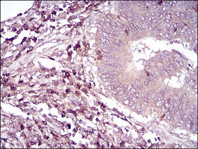
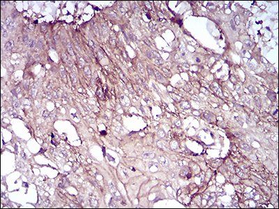
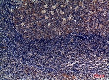
| Dilution range | Flow cytometry: Recommended dilution: 2 μg/ml. Immunohistochemistry (paraffin sections): Recommended dilution: 10 μg/ml; positive tissue: spleen. Western blotting: Non-reducing conditions. |
| Purity | Purified by protein-A affinity chromatography. |
| Conjugation | Unconjugated |
| Target | CD6 |
| Entrez | 923 |
| UniProt ID | P30203 |
| Storage | Store at 2-8°C. Do not freeze. |
| Buffer/Preservatives | Phosphate buffered saline (PBS), pH 7.4, 15 mM sodium azide |
| Alternative names | Anti-CD6 antibody Read more... |
| Note | For research use only |
| Application notes | Flow cytometry: Recommended dilution: 2 μg/ml. Immunohistochemistry (paraffin sections): Recommended dilution: 10 μg/ml; positive tissue: spleen. Western blotting: Recommended dilution: 1-2 μg/ml; non-reducing conditions. |
| Expiration Date | 12 months from date of receipt. |
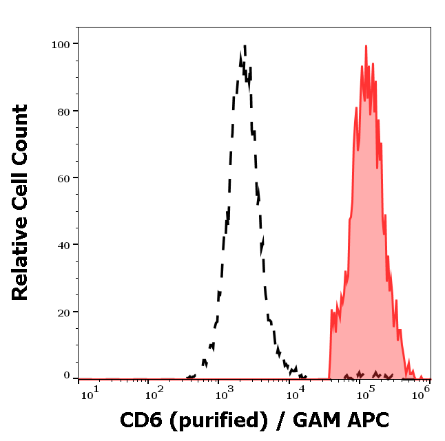
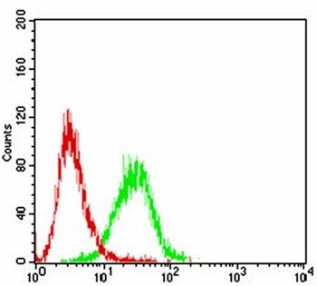
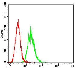
Separation of human CD6 positive lymphocytes (red-filled) from neutrophil granulocytes (black-dashed) in flow cytometry analysis (surface staining) of peripheral whole blood stained using anti-human CD6 (MEM-98) purified antibody (concentration in sample 2 μg/ml, GAM APC).
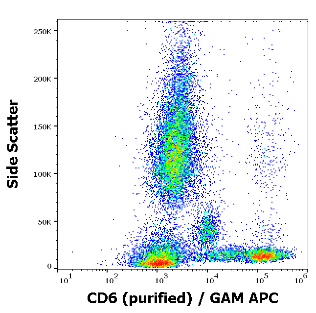
Flow cytometry surface staining pattern of human peripheral whole blood stained using anti-human CD6 (MEM-98) purified antibody (concentration in sample 2 μg/ml, GAM APC).
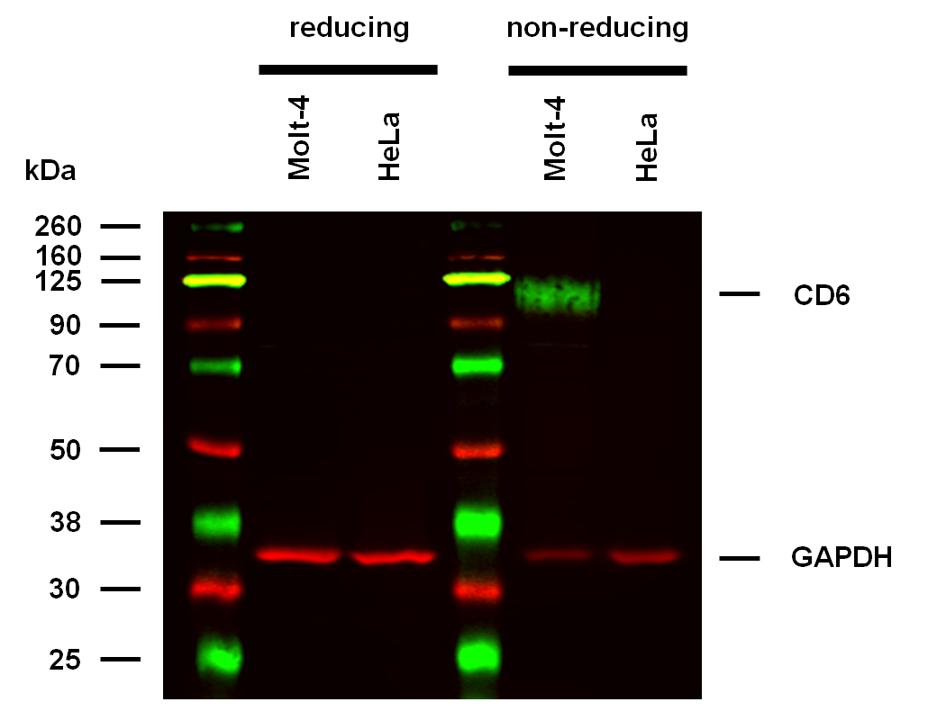
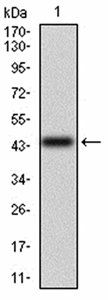
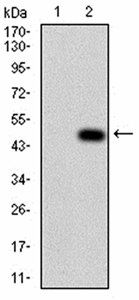
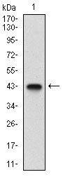
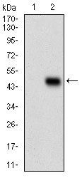
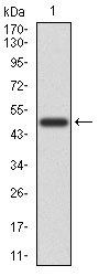
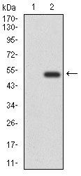
Anti-Hu CD6 Purified (clone MEM-98) works in WB application under non-reducing conditions. Western blotting analysis was performed on whole cell extracts (RIPA lysis buffer) of Molt-4 and HeLa cell lines, mixed and heated (100°C, 5 min) with reducing and non-reducing SDS-loading buffer. Samples were resolved using 10% Tris-glycine SDS gel electrophoresis. Nitrocellulose membrane blot was probed with mouse IgG1 monoclonal antibody MEM-98 (1 µg/ml), followed by IRDye 800CW Goat-anti-Mouse IgG (green). Mouse anti-GAPDH monoclonal antibody FF26A conjugated with DyLight 680 (0.1 µg/ml), was used as the loading control (red). Multiplex fluorescent Western blot detection was performed. CD6 molecules were detected at ~100 kDa in Molt-4 cell line under non-reducing conditions.
- Item 1 of 7
- Item 1 of 6
- Item 1 of 4
- Item 1 of 1
- Item 1 of 2
Submit a review
Filter by Rating

- 5 stars

- 4 stars
- 3 stars
- 2 stars
- 1 stars