You have no items in your shopping cart.
Cart summary
Item 1 of 3
Item 1 of 3
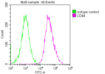
CD44 Antibody
Catalog Number: orb1252799

| Catalog Number | orb1252799 |
|---|---|
| Category | Antibodies |
| Description | CD44 Antibody |
| Species/Host | Mouse |
| Clonality | Monoclonal |
| Clone Number | 156-3C11 |
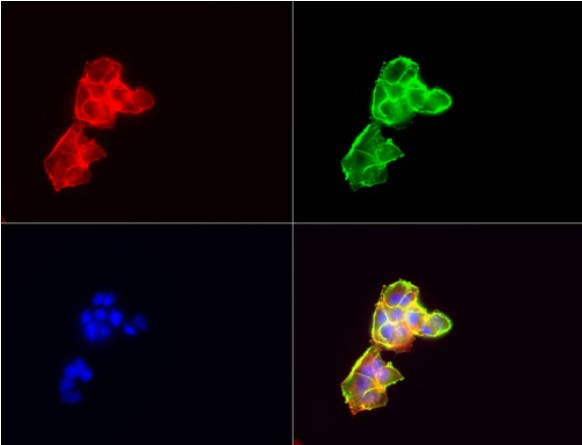
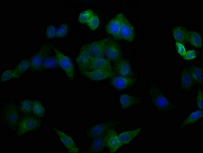
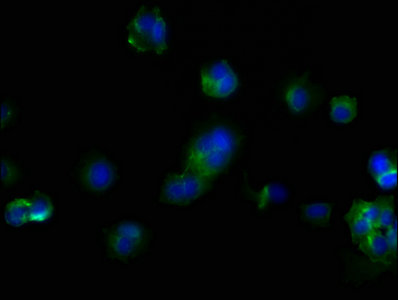
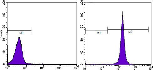
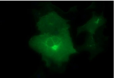
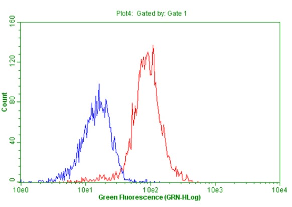
| Tested applications | ELISA, FC, IF, IHC, WB |
| Reactivity | Human, Monkey, Primate |
| Isotype | IgG2a, kappa |
| Immunogen | Stimulated human leukocytes were used as the immunogen for this CD44 / HCAM antibody. |
| Concentration | 0.2 mg/mL |
| Dilution range | ELISA: order BSA-free format for coatingFlow Cytometry: 0.5-1 ug/million cellsIF: 0.5-1 ug/mlWB: 0.5-1 ug/mlIHC (FFPE): 0.5-1 ug/ml for 30 minutes at RT (1)Prediluted format : incubate for 30 min at RT (2)The concentration stated for each application is a general starting point. Variations in protocols, secondaries and substrates may require the HCAM antibody to be titered up or down for optimal performance.1. Staining of formalin-fixed tissues requires boiling tissue sections in 10mM citrate buffer, pH 6.0, for 10-20 min followed by cooling at RT for 20 minutes.2. The prediluted format is supplied in a dropper bottle and is optimized for use in IHC. After epitope retrieval step (if required), drip mAb solution onto the tissue section and incubate at RT for 30 min. |
| Form/Appearance | Liquid |
| Conjugation | Unconjugated |
| Target | CD44 |
| UniProt ID | P16070 |
| Storage | Aliquot and Store at 2-8°C. Avoid freez-thaw cycles. |
| Buffer/Preservatives | PBS with 0.1 mg/ml rAlbumin and 0.05% sodium azide |
| Alternative names | CD44, CD44 molecule (Indian blood group), CDW44, C Read more... |
| Note | For research use only |
| Application notes | ELISA: order BSA-free format for coatingFlow Cytometry: 0.5-1 ug/million cellsIF: 0.5-1 ug/mlWB: 0.5-1 ug/mlIHC (FFPE): 0.5-1 ug/ml for 30 minutes at RT (1)Prediluted format : incubate for 30 min at RT (2)The concentration stated for each application is a general starting point. Variations in protocols, secondaries and substrates may require the HCAM antibody to be titered up or down for optimal performance.1. Staining of formalin-fixed tissues requires boiling tissue sections in 10mM citrate buffer, pH 6.0, for 10-20 min followed by cooling at RT for 20 minutes.2. The prediluted format is supplied in a dropper bottle and is optimized for use in IHC. After epitope retrieval step (if required), drip mAb solution onto the tissue section and incubate at RT for 30 min. |
| Expiration Date | 12 months from date of receipt. |
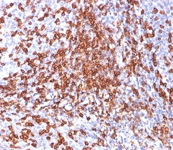
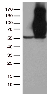
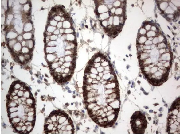
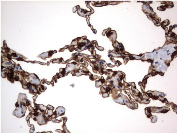
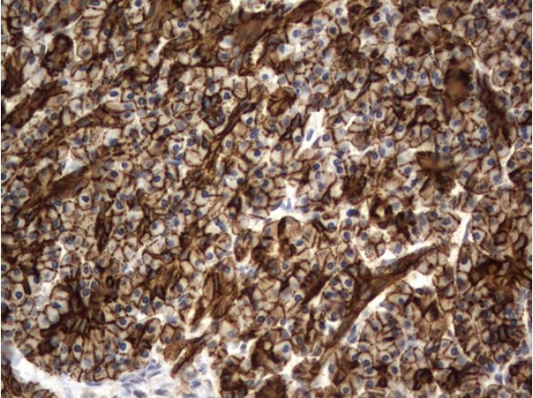
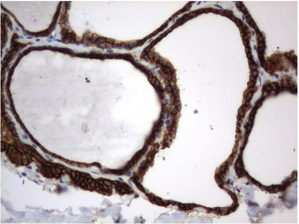
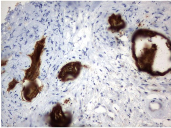
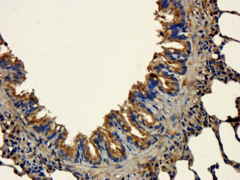
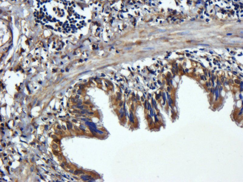
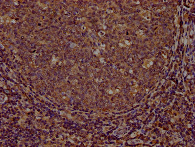
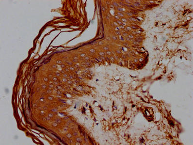
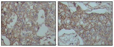
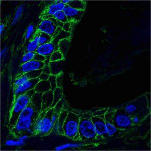
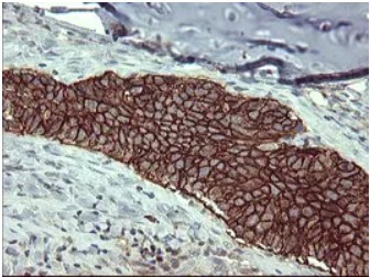
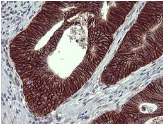
IHC testing of human tonsil stained with HCAM antibody (156-3C11).
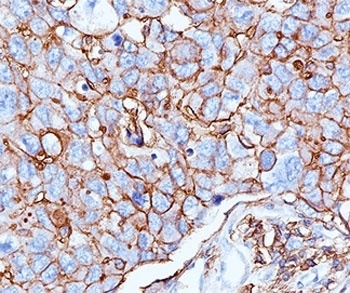
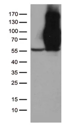
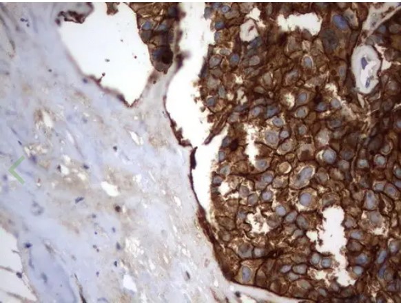
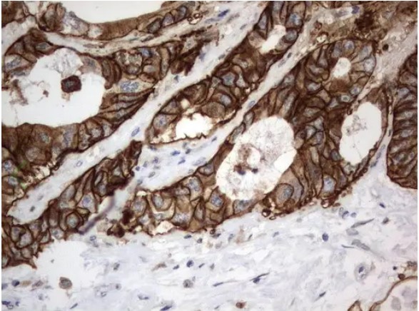
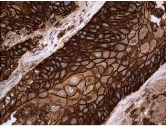
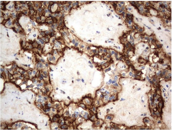
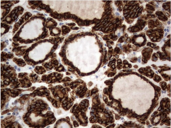
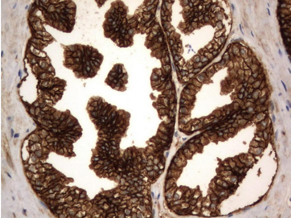
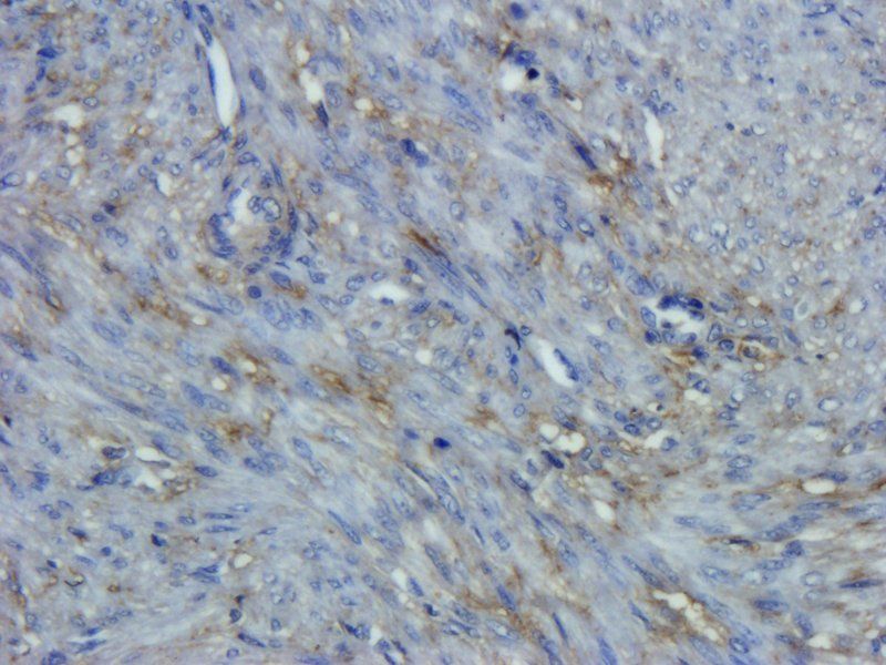
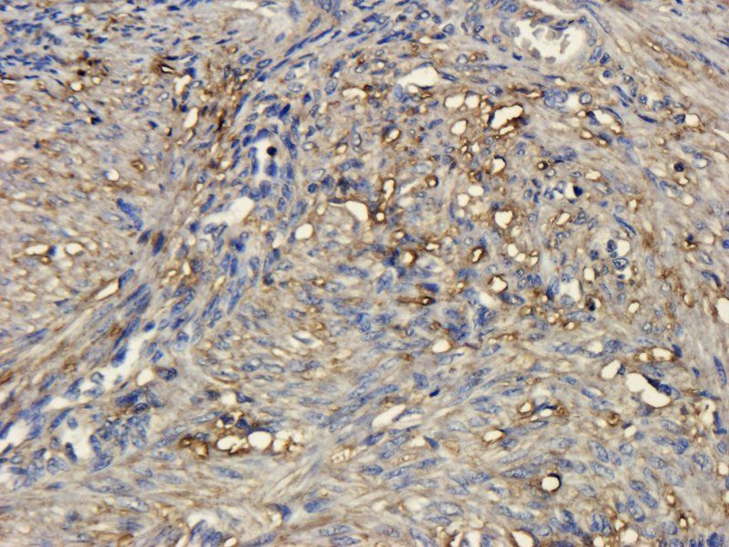
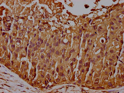
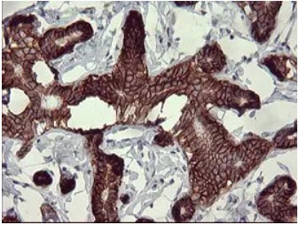
IHC testing of human breast cancer stained with HCAM antibody (156-3C11).
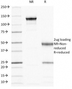
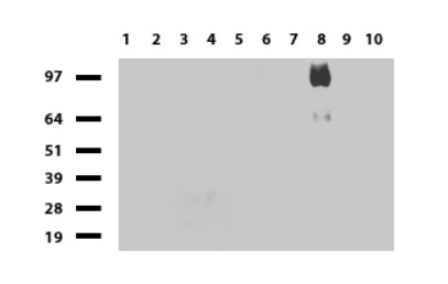
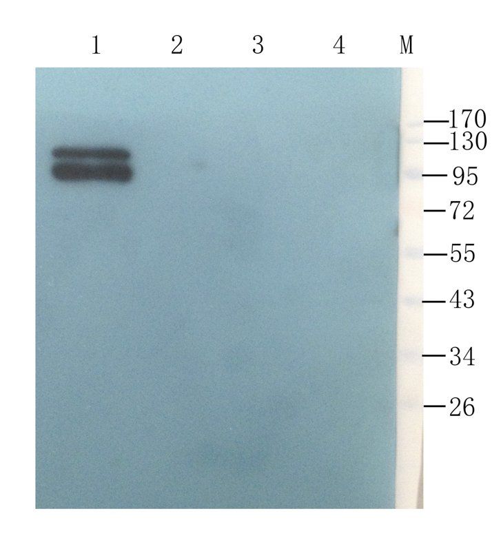
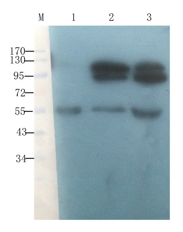
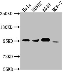
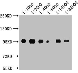
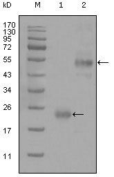
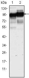
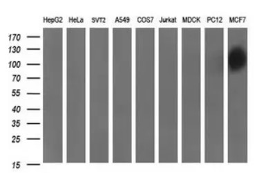
SDS-PAGE Analysis of Purified, BSA-Free HCAM Antibody (clone 156-3C11). Confirmation of Integrity and Purity of the Antibody.
- Item 1 of 16
- Item 1 of 9
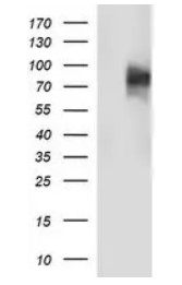
CD44 antibody [orb259645]
ELISA, IHC-P, WB
Human, Mouse, Porcine, Rat
Rabbit
Polyclonal
Unconjugated
100 μg, 200 μg - Item 1 of 8
- Item 1 of 7
- Item 1 of 8
Submit a review
Filter by Rating

- 5 stars

- 4 stars
- 3 stars
- 2 stars
- 1 stars
