You have no items in your shopping cart.
Cart summary
Item 1 of 16
Item 1 of 16
CD2AP Antibody (monoclonal, 5F8)
Catalog Number: orb507557
| Catalog Number | orb507557 |
|---|---|
| Category | Antibodies |
| Description | CD2AP Antibody (monoclonal, 5F8) |
| Species/Host | Mouse |
| Clonality | Monoclonal |
| Clone Number | 5F8 |
| Tested applications | FC, ICC, IF, IHC, WB |
| Reactivity | Human, Mouse, Rat |
| Isotype | Mouse IgG1 |
| Immunogen | E. coli-derived human CD2AP recombinant protein (Position: K253-K337). |
| Concentration | Adding 0.2 ml of distilled water will yield a concentration of 500 μg/ml. |
| Dilution range | Western blot, 0.1-0.5μg/ml Immunohistochemistry (Paraffin-embedded Section), 0.5-1μg/ml Immunocytochemistry/Immunofluorescence, 5 μg/ml Flow Cytometry, 1-3μg/1x106 cells |
| Form/Appearance | Lyophilized |
| Conjugation | Unconjugated |
| MW | 80 kDa |
| UniProt ID | Q9Y5K6 |
| Storage | Store at -20˚C for one year from date of receipt. After reconstitution, at 4˚C for one month. It can also be aliquotted and stored frozen at -20˚C for six months. Avoid repeated freeze-thaw cycles. |
| Alternative names | CD2-associated protein; Adapter protein CMS; Cas l Read more... |
| Note | For research use only |
| Expiration Date | 12 months from date of receipt. |

ICC analysis of CD2AP using anti-CD2AP antibody.CD2AP was detected in immunocytochemical section of A431 cell.Biotinylated goat anti-mouse IgG was used as secondary antibody.
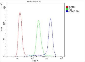
Flow Cytometry analysis of K562 cells using anti-D2AP antibody (Blue line).Isotype control antibody (Green line) was mouse IgG .Unlabelled sample (Red line) was also used as a control.
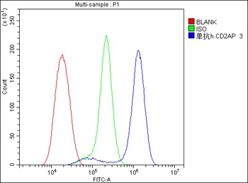
Flow Cytometry analysis of U20S cells using anti-CD2AP antibody (Blue line).Isotype control antibody (Green line) was rabbit IgG .Unlabelled sample (Red line) was also used as a control.
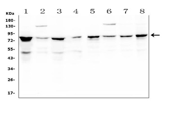
WB analysis of CD2AP using anti-CD2AP antibody.Lane 1:human K562 Cell;2:human A431 Cell;3:human HEK293 Cell;4:human U20S Cell;5:human HL-60 Cell;6:human MCF-7 Cell;7:human HeLa Cell;8:human PANC-1 Cell.
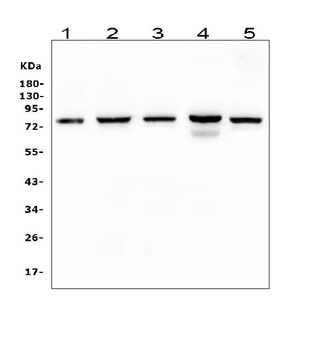
WB analysis of CD2AP using anti-CD2AP antibody.Lane 1:rat liver Tissue;2:rat testicular Tissue;3:mouse pancreas Tissue;4:mouse testicular Tissue;5:mouse RAW246.7 Cell.

IHC analysis of CD2AP using anti-CD2AP antibody.CD2AP was detected in paraffin-embedded section of human colon cancer.
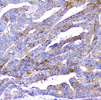
IHC analysis of CD2AP using anti-CD2AP antibody.CD2AP was detected in paraffin-embedded section of human colon cancer.

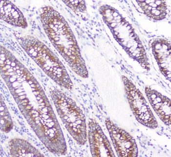
IHC analysis of CD2AP using anti-CD2AP antibody.CD2AP was detected in paraffin-embedded section of human placenta tissue.
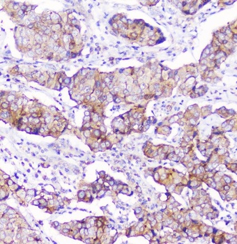
IHC analysis of CD2AP using anti-CD2AP antibody.CD2AP was detected in paraffin-embedded section of human mammary cancer.
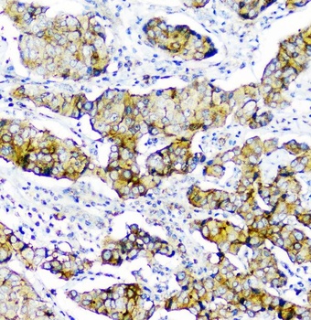
IHC analysis of CD2AP using anti-CD2AP antibody.CD2AP was detected in paraffin-embedded section of human mammary cancer.
IHC analysis of CD2AP using anti-CD2AP antibody.CD2AP was detected in paraffin-embedded section of human colon cancer.

IHC analysis of CD2AP using anti-CD2AP antibody.CD2AP was detected in paraffin-embedded section of human colon cancer.
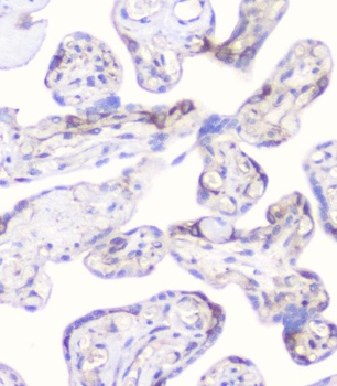
IHC analysis of CD2AP using anti-CD2AP antibody.CD2AP was detected in paraffin-embedded section of human placenta tissue.
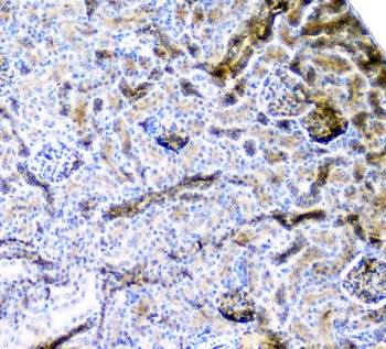
IHC analysis of CD2AP using anti-CD2AP antibody.CD2AP was detected in paraffin-embedded section of mouse kidney tissue.
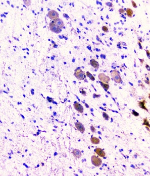
IHC analysis of CD2AP using anti-CD2AP antibody.CD2AP was detected in paraffin-embedded section of rat brain tissue.
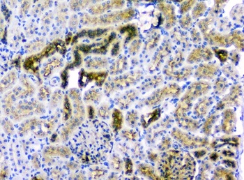
IHC analysis of CD2AP using anti-CD2AP antibody.CD2AP was detected in paraffin-embedded section of rat kidney tissue.
Submit a review
Filter by Rating

- 5 stars

- 4 stars
- 3 stars
- 2 stars
- 1 stars