You have no items in your shopping cart.
Cart summary
Item 1 of 7
Item 1 of 7
CARD6 Antibody
Catalog Number: orb1262399
| Catalog Number | orb1262399 |
|---|---|
| Category | Antibodies |
| Description | CARD6 Antibody |
| Species/Host | Rabbit |
| Clonality | Polyclonal |
| Tested applications | IHC-P, WB |
| Reactivity | Human |
| Isotype | Rabbit Ig |
| Immunogen | This CARD6 antibody is generated from rabbits immunized with a KLH conjugated synthetic peptide selected from the Center region of human CARD6. |
| Concentration | batch dependent |
| Dilution range | For IHC-P starting dilution is: 1:25For WB starting dilution is: 1:1000 |
| Form/Appearance | Liquid |
| Conjugation | Unconjugated |
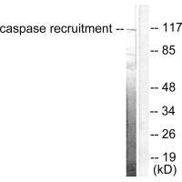
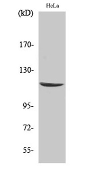
| MW | 116 kDa |
| Target | CARD6 |
| UniProt ID | Q9BX69 |
| NCBI | Q9BX69 |
| Storage | Store at 4°C for three months and -20°C, stable for up to one year. As with all antibodies care should be taken to avoid repeated freeze thaw cycles. Antibodies should not be exposed to prolonged high temperatures. |
| Buffer/Preservatives | Supplied in PBS with 0.09% (W/V) sodium azide. |
| Alternative names | Caspase recruitment domain-containing protein 6, C Read more... |
| Note | For research use only |
| Application notes | For IHC-P starting dilution is: 1:25For WB starting dilution is: 1:1000 |
| Expiration Date | 12 months from date of receipt. |
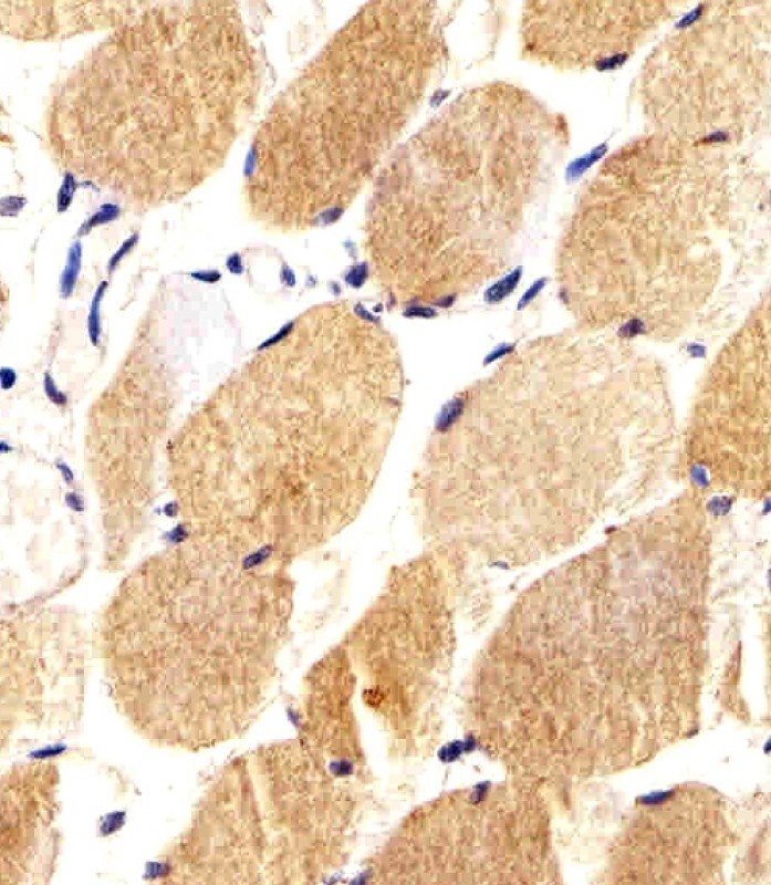
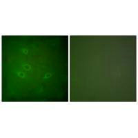
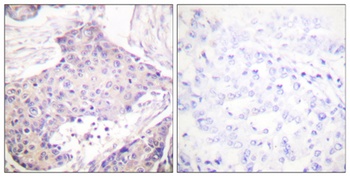
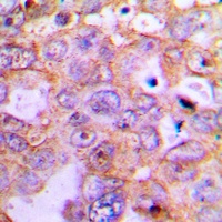
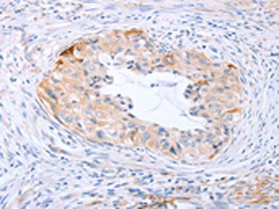
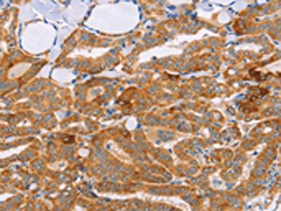
Immunohistochemical analysis of paraffin-embedded H. skeletal muscle section using CARD6 Antibody. Antibody was diluted at 1:25 dilution. A undiluted biotinylated goat polyvalent antibody was used as the secondary, followed by DAB staining.
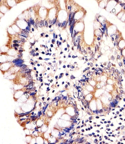
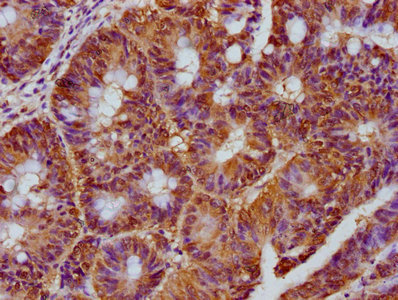
Immunohistochemical analysis of paraffin-embedded H. colon section using CARD6 Antibody. Antibody was diluted at 1:25 dilution. A undiluted biotinylated goat polyvalent antibody was used as the secondary, followed by DAB staining.
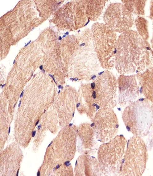
Immunohistochemical analysis of paraffin-embedded H. skeletal muscle section using CARD6 Antibody. Antibody was diluted at 1:25 dilution. A undiluted biotinylated goat polyvalent antibody was used as the secondary, followed by DAB staining.
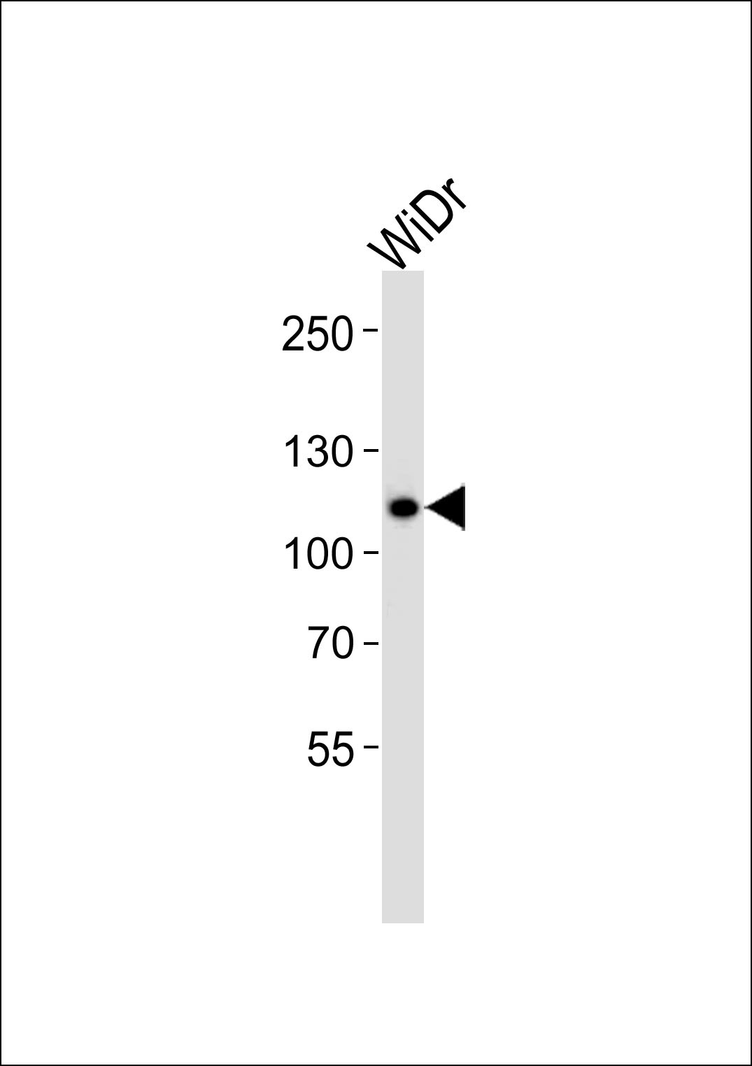
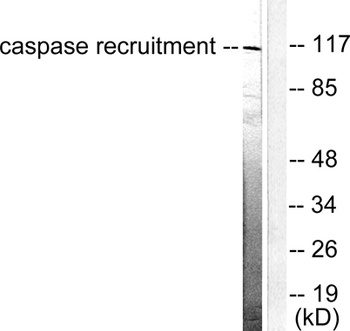
Western blot analysis of lysate from WiDr cell line, using CARD6 Antibody at 1:1000.
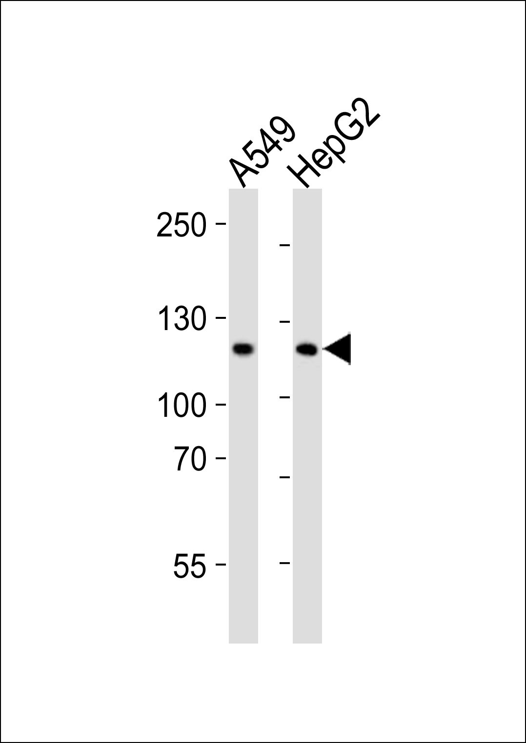
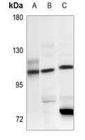
Western blot analysis of lysates from A549, HepG2 cell line (from left to right), using CARD6 Antibody at 1:1000 at each lane.
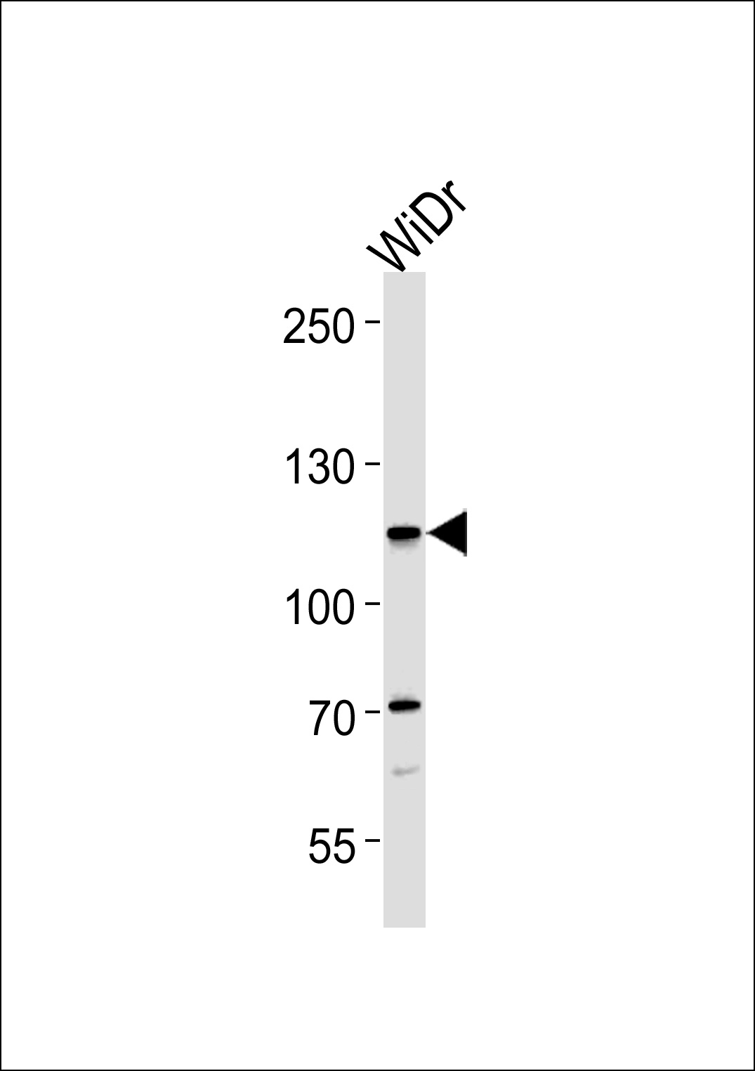
Western blot analysis in WiDr cell line lysates (35 ug/lane).
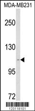
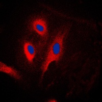
Western blot analysis of CARD6 Antibody in MDA-MB231 cell line lysates (35 ug/lane)
- Item 1 of 3
- Item 1 of 4
- Item 1 of 3
- Item 1 of 2
- Item 1 of 2

Submit a review
Filter by Rating

- 5 stars

- 4 stars
- 3 stars
- 2 stars
- 1 stars