You have no items in your shopping cart.
Cart summary
Item 1 of 3
Item 1 of 3
Calponin 1 antibody (MSVA-455R)
Catalog Number: orb2276529

| Catalog Number | orb2276529 |
|---|---|
| Category | Antibodies |
| Description | Calponin 1 antibody validated for immunohistochemistry on 76 different Normal Tissues |
| Species/Host | Rabbit |
| Clonality | Monoclonal |
| Clone Number | Biorbyt-455R |
| Tested applications | IHC |
| Reactivity | Human |
| Isotype | IgG |
| Immunogen | Synthetic peptide corresponding to residues on the C-terminus of human Calponin was used as an immunogen. |
| Dilution range | 1:100-1:200 |
| Conjugation | Unconjugated |
| UniProt ID | P51911 |
| Storage | Antibody with azide – store at 2 to 8°C. Antibody without azide – store at -20 to -80°C. Antibody is stable for 24 months. Non- hazardous. No MSD required. |
| Alternative names | Calponin 1 basic smooth muscle; Calponin H1 smooth Read more... |
| Note | For research use only |
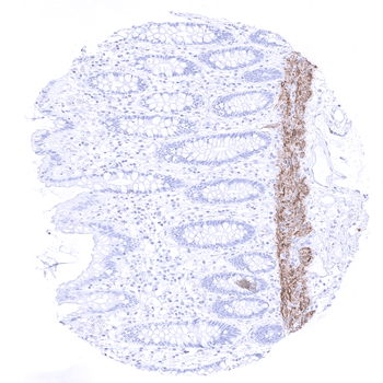
| Application notes | Positive Control: Colon: A moderate to strong Calponin 1 staining should be seen in the muscularis mucosae, muscularis propria and in the walls of larger blood vessels. Negative Control: Colon: Calponin 1 staining should be absent in all epithelial cells. Cellular Localization: Cytoplasmic Protocol Recommendations: Manual Protocol: Freshly cut sections should be used (less than 10 days between cutting and staining). Heat-induced antigen retrieval for 5 minutes in an autoclave at 121°C in pH 7,8 Target Retrieval Solution buffer. Apply the antibody at a dilution of 1:150 at 37°C for 60 minutes. Visualization of bound antibody by the EnVision Kit (Dako, Agilent) according to the manufacturer’s directions. |
| Expiration Date | 12 months from date of receipt. |

A moderate intensity Calponin 1 immunostaining is seen in the muscularis mucosae in a colon sample.
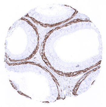
A strong calponin 1 immunostaining occurs in the periductal smooth muscle layer in the epididymis.

Calponin 1 immunostaining is regularly seen in myoepithelial cells of the breast.
- Item 1 of 3
- Item 1 of 3