You have no items in your shopping cart.
Cart summary
Item 1 of 7
Item 1 of 7
CAD Antibody
Catalog Number: orb1270371

| Catalog Number | orb1270371 |
|---|---|
| Category | Antibodies |
| Description | CAD Antibody |
| Target | CAD |
| Clonality | Polyclonal |
| Isotype | Rabbit Ig |
| Conjugation | Unconjugated |
| Reactivity | Human |
| Predicted Reactivity | Mouse |
| Form/Appearance | Liquid |
| Concentration | batch dependent |
| Buffer/Preservatives | Supplied in PBS with 0.09% (W/V) sodium azide. |
| Immunogen | This CAD antibody is generated from rabbits immunized with a KLH conjugated synthetic peptide between 780-809 amino acids from the Central region of human CAD. |
| UniProt ID | P27708 |
| MW | 243 kDa |
| Tested applications | FC, IHC-P, WB |
| Application notes | For FACS starting dilution is: 1:25For IHC-P starting dilution is: 1:25For WB starting dilution is: 1:2000 |
| Antibody Type | Primary Antibody |
| Storage | Maintain refrigerated at 2-8°C for up to 2 weeks. For long term storage store at -20°C in small aliquots to prevent freeze-thaw cycles. |
| Alternative names | CAD protein, Glutamine-dependent carbamoyl-phospha Read more... |
| Note | For research use only |
| NCBI | P27708 |
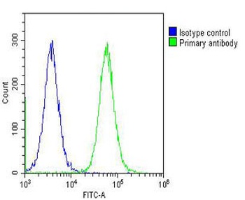
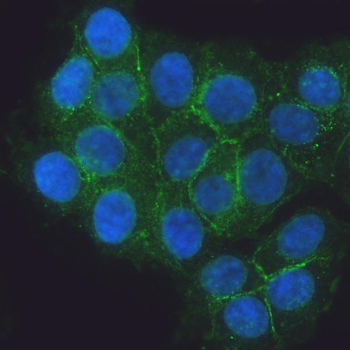
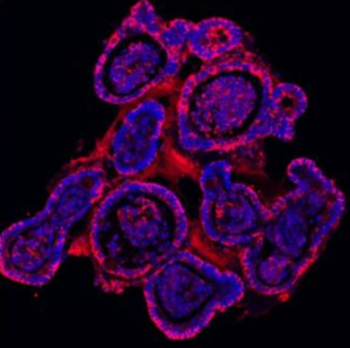
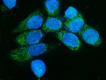
Overlay histogram showing Hela cells stained with Antibody (green line). The cells were fixed with 2% paraformaldehyde (10 min) and then permeabilized with 90% methanol for 10 min. The cells were then icubated in 2% bovine serum albumin to block non-specific protein-protein interactions followed by the antibody (1:25 dilution) for 60 min at 37oC. The secondary antibody used was Goat-Anti-Rabbit IgG, DyLight 488 Conjugated Highly Cross-Adsorbed (OH191631) at 1/200 dilution for 40 min at 37oC. Isotype control antibody (blue line) was rabbit IgG (1 ug/1x10^6 cells) used under the same conditions. Acquisition of >10000 events was performed.
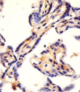
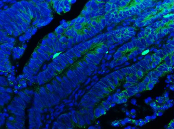
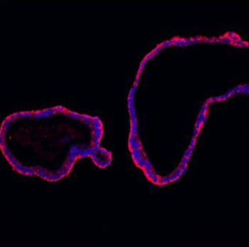
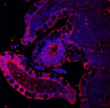
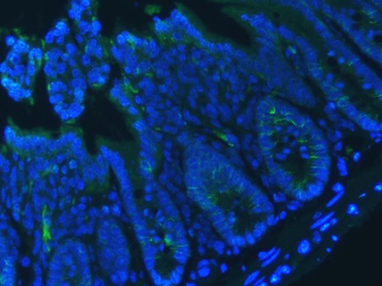
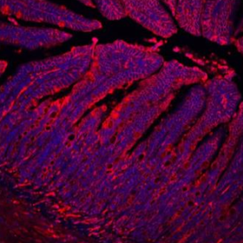
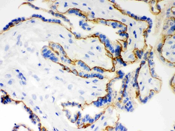
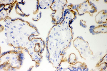
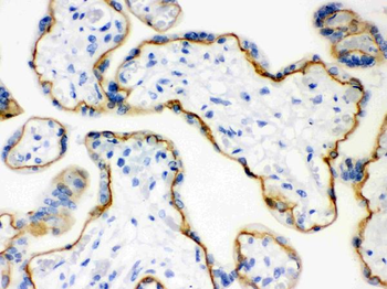
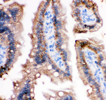
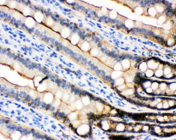
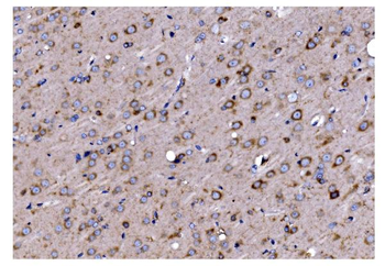
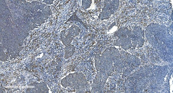
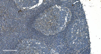
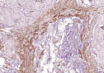
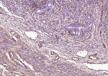
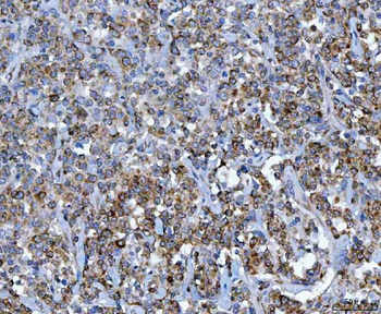
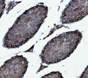
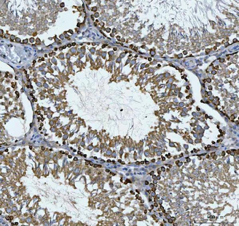
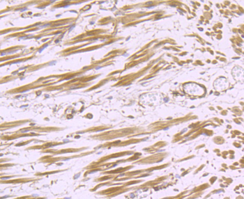
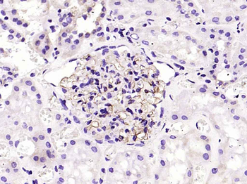
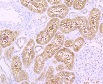
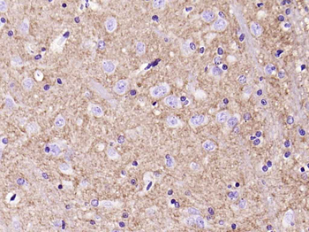
Antibody staining CAD in human placenta tissue sections by Immunohistochemistry (IHC-P - paraformaldehyde-fixed, paraffin-embedded sections).
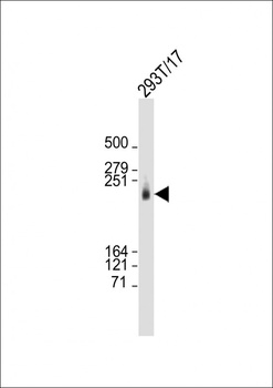
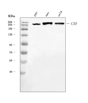
Western Blot at 1:2000 dilution + 293T/17 whole cell lysate Lysates/proteins at 20 ug per lane.
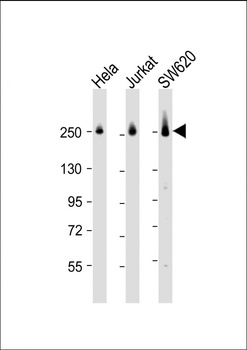
Western Blot at 1:2000 dilution Lane 1: Hela whole cell lysate Lane 2: Jurkat whole cell lysate Lane 3: SW620 whole cell lysate Lysates/proteins at 20 ug per lane.
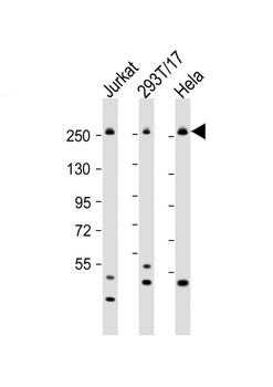
Western Blot at 1:2000 dilution Lane 1: Jurkat whole cell lysate Lane 2: 293T/17 whole cell lysate Lane 3: Hela whole cell lysate Lysates/proteins at 20 ug per lane.
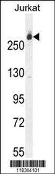
Western blot analysis in Jurkat cell line lysates (35 ug/lane).
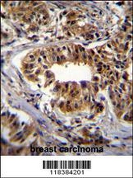
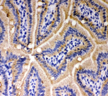
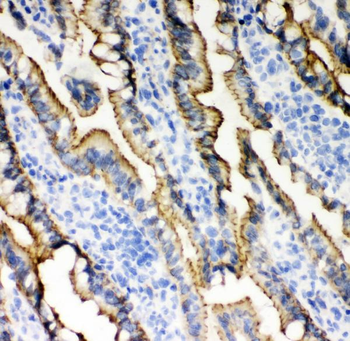
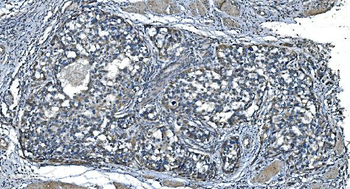
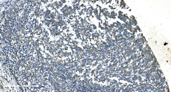
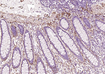
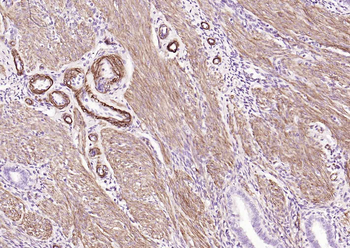
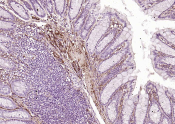
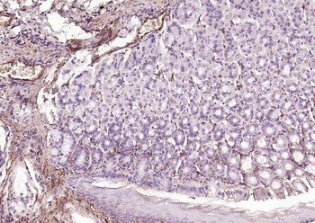
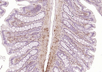
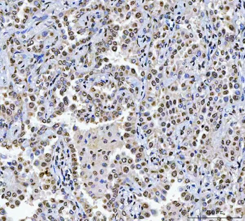
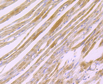
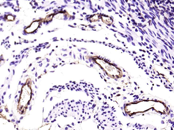
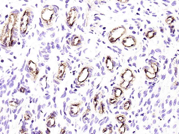
CAD Antibody immunohistochemistry analysis in formalin fixed and paraffin embedded human breast carcinoma followed by peroxidase conjugation of the secondary antibody and DAB staining.
- Item 1 of 15
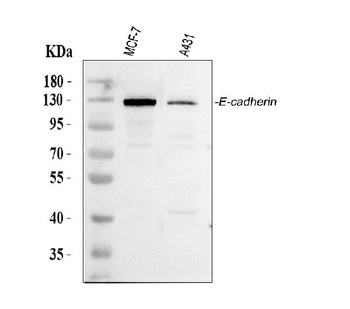
Anti-E Cadherin 1/CDH1 Antibody [orb308856]
ELISA, ICC, IF, IHC, IHC-Fr, WB
Human, Mouse, Rat
Rabbit
Polyclonal
Unconjugated
10 μg, 100 μg - Item 1 of 11
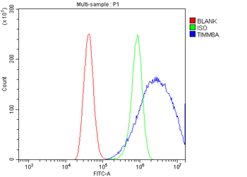
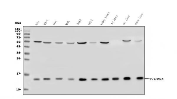
Anti-TIMM8A/DDP Antibody [orb670328]
ELISA, ICC, IF, IHC, WB
Human, Monkey, Mouse, Rat
Rabbit
Polyclonal
Unconjugated
10 μg, 100 μg - Item 1 of 10
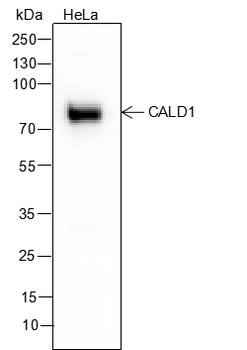
Caldesmon Recombinant Mouse Monoclonal Antibody [orb1173066]
IF, IHC-Fr, IHC-P, WB
Human, Mouse, Rat
Human, Mouse, Rat
Mouse
Recombinant
Unconjugated
25 μl, 100 μl, 50 μl - Item 1 of 9
Anti-CAD Antibody [orb1290024]
ELISA, ICC, IF, IHC, WB
Human, Mouse, Rat
Rabbit
Polyclonal
Unconjugated
10 μg, 100 μg - Item 1 of 9
H Cadherin Recombinant Rabbit Monoclonal Antibody [orb704170]
IF, IHC-Fr, IHC-P
Mouse, Rat
Human, Mouse, Rat
Rabbit
Recombinant
Unconjugated
100 μl, 50 μl