You have no items in your shopping cart.
Cart summary
Item 1 of 1
Blood Group Lewis b Antibody / FUT3
Catalog Number: orb385735

| Catalog Number | orb385735 |
|---|---|
| Category | Antibodies |
| Description | The Lewis histo-blood group system comprises a set of fucosylated glycosphingolipids that are synthesized by exocrine epithelial cells and circulate in body fluids. The glycosphingolipids function in embryogenesis, tissue differentiation, tumor metastasis, inflammation, and bacterial adhesion. They are secondarily absorbed to red blood cells giving rise to their Lewis phenotype. This gene is a member of the fucosyltransferase family, which catalyzes the addition of fucose to precursor polysaccharides in the last step of Lewis antigen biosynthesis. It encodes an enzyme with alpha(1,3)-fucosyltransferase and alpha(1,4)-fucosyltransferase activities. Lewis blood group antigens are carbohydrate moieties structurally integrated in mucous secretions. Lewis antigen system alterations have been described in gastric carcinoma and associated lesions. Anomalous expression of Lewis B antigen has been found in some non-secretory gastric carcinomas and colorectal cancers. |
| Species/Host | Mouse |
| Clonality | Monoclonal |
| Clone Number | 2-25LE or LWB01 |
| Tested applications | IHC-P |
| Reactivity | Human |
| Isotype | Mouse IgG1, kappa |
| Immunogen | Mucin isolated from human ovarian cyst fluid was used as the immunogen for this Lewis b antibody. |
| Antibody Type | Primary Antibody |
| Dilution range | Immunohistochemistry (FFPE): 1-2ug/ml for 30 min at RT |
| Purity | Protein G affinity chromatography |
| Conjugation | Unconjugated |
| Formula | 0.2 mg/ml in 1X PBS with 0.1 mg/ml BSA (US sourced) and 0.05% sodium azide |
| Hazard Information | This Lewis b antibody is available for research use only. |
| UniProt ID | P21217 |
| Storage | Store the Lewis b antibody at 2-8°C (with azide) or aliquot and store at -20°C or colder (without azide). |
| Buffer/Preservatives | 0.2 mg/ml in 1X PBS with 0.1 mg/ml rAlbumin (US sourced) and 0.05% sodium azide |
| Note | For research use only |
| Application notes | The optimal dilution of the Blood Group Lewis b antibody for each application should be determined by the researcher.1. Staining of formalin-fixed tissues is enhanced by boiling tissue sections in pH 9 10mM Tris with 1mM EDTA for 10-20 min followed by cooling at RT for 20 minutes.2. The prediluted format is supplied in a dropper bottle and is optimized for use in IHC. After epitope retrieval step (if required), drip mAb solution onto the tissue section and incubate at RT for 30 min. |
| Expiration Date | 12 months from date of receipt. |

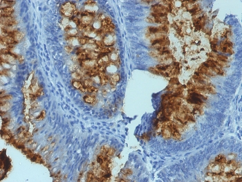
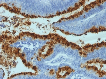
IHC staining of FFPE human colon carcinoma with FUT3 antibody (clone SPM194).
- Item 1 of 1
- Item 1 of 1
- Item 1 of 1