You have no items in your shopping cart.
Cart summary
Item 1 of 1
Beta galactosidase antibody (Biotin)
Catalog Number: orb344931
| Catalog Number | orb344931 |
|---|---|
| Category | Antibodies |
| Description | Beta galactosidase antibody (Biotin) |
| Species/Host | Rabbit |
| Clonality | Polyclonal |
| Tested applications | ELISA, IHC, WB |
| Isotype | IgG |
| Immunogen | Beta Galactosidase (E.coli) |
| Concentration | 1.0 mg/mL |
| Dilution range | ELISA: 1:10,000 - 1:50,000, IHC: 1:1,000 - 1:5,000, WB: 1:2,000 - 1:10,000 |
| Form/Appearance | Liquid (sterile filtered) |
| Purity | Anti-BETA GALACTOSIDASE Antibody, Biotin Conjugated, is an IgG fraction antibody purified from monospecific antiserum by a multi-step process which includes delipidation, salt fractionation and ion exchange chromatography followed by extensive dialysis against the buffer stated above. Assay by immunoelectrophoresis resulted in a single precipitin arc against anti-Biotin, anti-Rabbit Serum as well as purified and partially purified beta-Galactosidase [E.coli]. Cross reactivity against β-Galactosidase from other sources may occur but have not been specifically determined. |
| Conjugation | Biotin |
| UniProt ID | P00722 |
| NCBI | NP_414878.1 |
| Storage | Store vial at -20° C or below prior to opening. This vial contains a relatively low volume of reagent (25 µL). To minimize loss of volume dilute 1:10 by adding 225 µL of the buffer stated above directly to the vial. Recap, mix thoroughly and briefly centrifuge to collect the volume at the bottom of the vial. Use this intermediate dilution when calculating final dilutions as recommended below. Store the vial at -20°C or below after dilution. Avoid cycles of freezing and thawing. |
| Buffer/Preservatives | 0.01% (w/v) Sodium Azide |
| Alternative names | rabbit anti Beta Galactosidase Antibody Biotin Con Read more... |
| Note | For research use only |
| Application notes | Anti-Beta Galactosidase Biotin Conjugated Antibody has been tested by Western blot and is suitable for ELISA, immunohistochemistry, immunomicroscopy as well as other antibody based assays using streptavidin or avidin conjugates requiring lot-to-lot consistency. The antibody recognizes both frozen tissue sections, paraffin embedded tissue and 4% paraformaldehyde fixed tissue for most immunohistochemical analysis. A 1:5,000 dilution has been reported to be successful for staining by immunoblot of beta-galactosidase fusion proteins after transfer using a semi-dry transfer apparatus. A 1:1,500 dilution has been reported to detect beta-galactosidase in adult rat spinal cord tissue after infection with helper-dependent adenovirus expressing lacZ. In this particular experiment, tissue was perfused with 4% paraformaldehyde and cryostat-cut (35 µm) to produce free-floating sections. A 1:5,000 dilution has been reported to be successful for staining of brain sections from transgenic mice expressing nuclear beta-galactosidase when assayed by immunofluorescence microscopy. A 1:5,000 dilution has been reported for immunofluorescent staining of methanol fixed, devitellinized Drosophila embryos. Although a wide range of conditions was reported to be effective, a 1:10,000 dilution was noted to show no background and to be suitable for double labeling experiments. Optimal titers for other applications should be determined by the researcher. |
| Expiration Date | 12 months from date of receipt. |
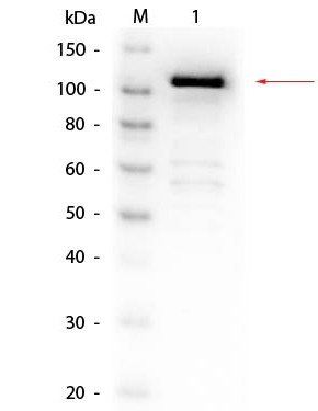
Western blot analysis of Lane 1: Beta Galactosidase. Load: 50 ng per lane using Beta Galactosidase antibody (Biotin)
Submit a review
Filter by Rating

- 5 stars

- 4 stars
- 3 stars
- 2 stars
- 1 stars