You have no items in your shopping cart.
Cart summary
Item 1 of 4
Item 1 of 4
Beta-actin Antibody (Ascites)
Catalog Number: orb2649354

| Catalog Number | orb2649354 |
|---|---|
| Category | Antibodies |
| Description | Mouse Monoclonal Antibody (Mab) |
| Species/Host | Mouse |
| Clonality | Monoclonal |
| Clone Number | 8H10D10 |
| Tested applications | IHC-P, WB |
| Reactivity | Human |
| Isotype | IgG |
| Antibody Type | Primary Antibody |
| Dilution range | WB: 1:1000, WB: 1:5000~20000, IHC-P: 1:10~50, IHC-P: 1:10~50 |
| Form/Appearance | Mouse monoclonal antibody supplied in crude ascites with 0.09% (W/V) sodium azide. |
| Conjugation | Unconjugated |
| MW | 41737 Da |
| Target | ACTB recombinant protein is used to produce this monoclonal antibody. |
| UniProt ID | P60709 |
| NCBI | NP_001092.1 |
| Storage | Maintain refrigerated at 2-8°C for up to 2 weeks. For long term storage store at -20°C in small aliquots to prevent freeze-thaw cycles. |
| Alternative names | Actin, cytoplasmic 1, Beta-actin, Actin, cytoplasm Read more... |
| Note | For research use only |
| Expiration Date | 12 months from date of receipt. |

All lanes: Anti-Beta-actin Antibody at 1:1000 dilution. Lane 1: Hela whole cell lysate. Lane 2: NIH/3T3 whole cell lysate. Lane 3: HepG2 whole cell lysate. Lane 4: MCF-7 whole cell lysate. Lane 5: A431 whole cell lysate. Lysates/proteins at 20 µg per lane. Secondary: Goat Anti-Mouse IgG, (H+L), Peroxidase conjugated at 1/8000 dilution.Observed band size: 42 KDa. Blocking/Dilution buffer: 5% NFDM/TBST.
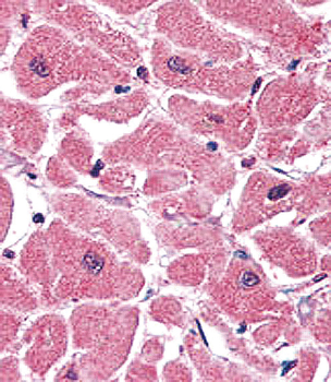
Formalin-fixed and paraffin-embedded human heart tissue reacted with Beta-actin Monoclonal Antibody, which was peroxidase-conjugated to the secondary antibody, followed by AEC staining. This data demonstrates the use of this antibody for immunohistochemistry; clinical relevance has not been evaluated.
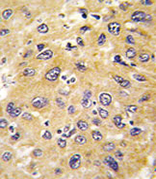
Formalin-fixed and paraffin-embedded human hepatocarcinoma tissue reacted with Beta-actin Monoclonal Antibody, which was peroxidase-conjugated to the secondary antibody, followed by DAB staining. This data demonstrates the use of this antibody for immunohistochemistry; clinical relevance has not been evaluated.
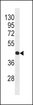
Western blot analysis of anti-Beta-actin Monoclonal Antibody in HL-60 cell line lysates (35 μg/lane). Beta-actin (arrow) was detected using the purified Mab.

Anti-Beta-Actin Monoclonal Loading Control [orb1675316]
ELISA, WB
Human
Mouse
Monoclonal
Unconjugated
1 mg
