You have no items in your shopping cart.
Cart summary
Item 1 of 6
Item 1 of 6
ATP5B Antibody (Center)
Catalog Number: orb1788259

| Catalog Number | orb1788259 |
|---|---|
| Category | Antibodies |
| Description | Affinity Purified Rabbit Polyclonal Antibody (Pab) |
| Target | This ATP5B antibody is generated from rabbits immunized with a KLH conjugated synthetic peptide between 135-163 amino acids from the Central region of human ATP5B. |
| Clonality | Polyclonal |
| Species/Host | Rabbit |
| Isotype | Rabbit IgG |
| Conjugation | Unconjugated |
| Reactivity | Human, Mouse, Rat |
| Form/Appearance | Purified polyclonal antibody supplied in PBS with 0.09% (W/V) sodium azide. This antibody is purified through a protein A column, followed by peptide affinity purification. |
| Immunogen | 135-163 aa |
| UniProt ID | P06576 |
| MW | 56560 Da |
| Tested applications | FC, IF, IHC-P, WB |
| Dilution range | IF: 1:25, WB: 1:1000, IHC-P: 1:25, IHC-P: 1:25, IHC-P: 1:50~100, FC: 1:10~50 |
| Antibody Type | Primary Antibody |
| Storage | Maintain refrigerated at 2-8°C for up to 2 weeks. For long term storage store at -20°C in small aliquots to prevent freeze-thaw cycles |
| Alternative names | ATP5B; ATPMB; ATPSB; ATP synthase subunit beta, mi Read more... |
| Note | For research use only |
| NCBI | NP_001677.2 |
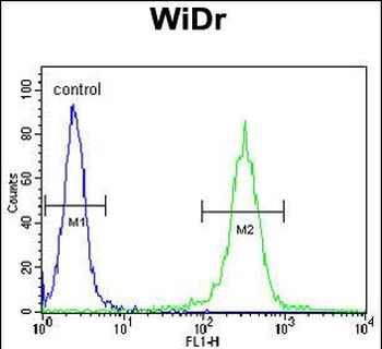
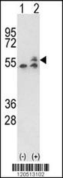
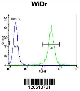
ATP5B Antibody (Center) flow cytometric analysis of WiDr cells (right histogram) compared to a negative control cell (left histogram). FITC-conjugated goat-anti-rabbit secondary antibodies were used for the analysis.

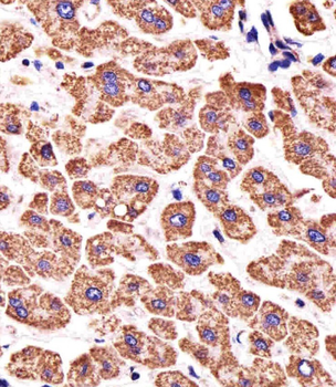
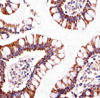
Immunohistochemical analysis of paraffin-embedded H. liver section using ATP5B Antibody (Center). Diluted at 1:25 dilution. A undiluted biotinylated goat polyvalent antibody was used as the secondary, followed by DAB staining.
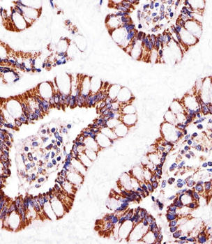
Immunohistochemical analysis of paraffin-embedded H. small intestine section using ATP5B Antibody (Center). Diluted at 1:25 dilution. A undiluted biotinylated goat polyvalent antibody was used as the secondary, followed by DAB staining.
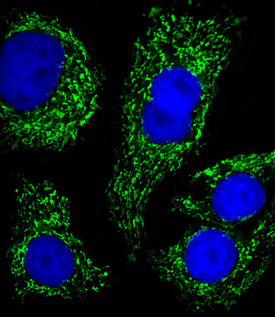
Fluorescent image of SK-BR-3 cells stained with ATP5B Antibody (Center). Diluted at 1:25 dilution. An Alexa Fluor 488-conjugated goat anti-rabbit lgG at 1:400 dilution was used as the secondary antibody (green). DAPI was used to stain the cell nuclear (blue).
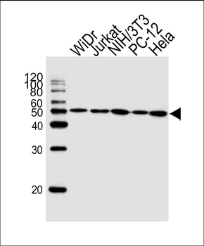
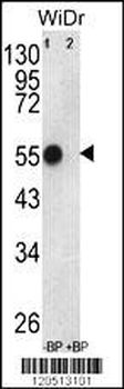
Western blot analysis of lysates from WiDr, Jurkat, mouse NIH/3T3, rat PC-12, Hela cell line (from left to right), using ATP5B Antibody (Center). Diluted at 1:1000 at each lane. A goat anti-rabbit IgG H&L (HRP) at 1:10000 dilution was used as the secondary antibody.
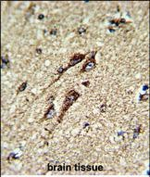
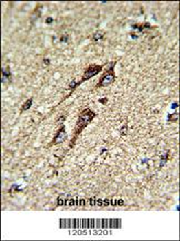
Formalin-fixed and paraffin-embedded human brain tissue reacted with ATP5B Antibody (Center), which was peroxidase-conjugated to the secondary antibody, followed by DAB staining. This data demonstrates the use of this antibody for immunohistochemistry; clinical relevance has not been evaluated.
- Item 1 of 7
ATP5B Antibody (Center) [orb1931307]
FC, IF, IHC-P, WB
Mouse, Rat
Human
Rabbit
Polyclonal
Unconjugated
100 μl, 50 μl