You have no items in your shopping cart.
Cart summary
Item 1 of 2
Item 1 of 2
Anti-HLA-G Purified Azide Free
Catalog Number: orb44434

| Catalog Number | orb44434 |
|---|---|
| Category | Antibodies |
| Description | Mouse Monoclonal to HLA-G. |
| Clonality | Monoclonal |
| Clone Number | MEM-G/9 |
| Tested applications | ELISA, FC, ICC, IHC-Fr, IP |
| Reactivity | Human |
| Isotype | Mouse IgG1 |
| Immunogen | Recombinant human HLA-G refolded with beta2-microglobulin and peptide. |
| Antibody Type | Primary Antibody |
| Concentration | 1 mg/ml |
| Dilution range | Flow cytometry: Recommended dilution: 1-5 μg/ml; positive control: JEG-3 human choriocarcinoma cell line. Immunocytochemistry: Recommended dilution: 2-5 μg/ml. For fixation details see: Emadi et al., Biotech Histochem. 2022 Feb;97(2):136-142.ELISA: The antibody MEM-G/9 has been tested as the capture antibody in a sandwich ELISA for analysis of human HLA-G in combination with antibody B2M-01 or with antibody W6/32. Coating antibody 10 μg/ml, detection antibody (biotin or peroxidase conjugate) 1 μg/ml. Immunohistochemistry: Recommended dilution: 5-10 μg/ml. |
| Purity | Purified by protein-A affinity chromatography. |
| Conjugation | Unconjugated |
| Target | HLA-G |
| Entrez | 3135 |
| UniProt ID | P17693 |
| RRID | AB_10995799 |
| Storage | Maintain refrigerated at 2-8°C for up to 2 weeks. For long term storage store at -20°C in small aliquots to prevent freeze-thaw cycles. |
| Buffer/Preservatives | Phosphate buffered saline (PBS), pH 7.4 |
| Alternative names | Anti-HLA-G antibody Read more... |
| Note | For research use only |
| Application notes | Flow cytometry: Recommended dilution: 1-5 μg/ml; positive control: JEG-3 human choriocarcinoma cell line. Immunocytochemistry: Recommended dilution: 2-5 μg/ml. ELISA: The antibody MEM-G/9 has been tested as the capture antibody in a sandwich ELISA for analysis of human HLA-G in combination with antibody B2M-01 or with antibody W6/32. Coating antibody 10 μg/ml, detection antibody (biotin or peroxidase conjugate) 1 μg/ml. Immunohistochemistry: Recommended dilution: 5-10 μg/ml. |
| Expiration Date | 12 months from date of receipt. |

Separation of HLA-G trasnfected LCL cells (red-filled) from K562 cells (black-dashed) in flow cytometry analysis (surface staining) stained using anti-human HLA-G (MEM-G/9) purified antibody (concentration in sample 0.3 μg/ml, GAM APC).
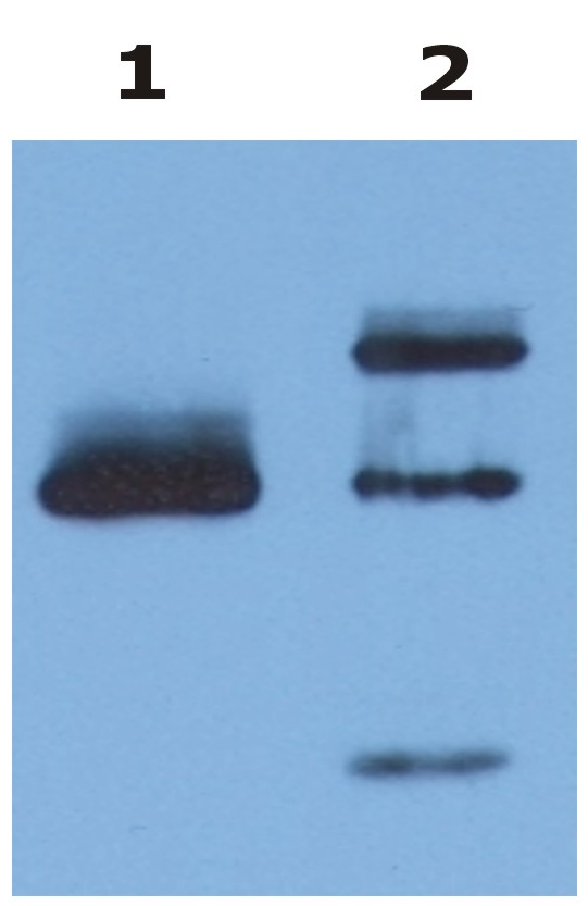
Immunoprecipitation of HLA-G from HLA-G1 transfectants (LCL-HLA-G1) by anti-human HLA-G (MEM-G/9) and protein G. HLA-G was detected by anti-human HLA-G (clone 4H84) and goat anti-mouse HRP in cell lysate (Lane 1) and in the immunoprecipitate (Lane 2).
